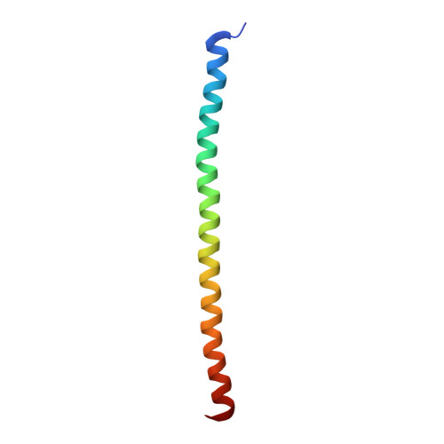
3HRN

crystal structure of a C-terminal coiled coil domain of Transient receptor potential (TRP) channel subfamily P member 2 (TRPP2, polycystic kidney disease 2)


Experimental Data Snapshot

  • Method: X-RAY DIFFRACTION
  • Resolution: 1.90 Å
  • R-Value Free: 0.260 
  • R-Value Work: 0.199 
  • R-Value Observed: 0.205 

Starting Model: experimental
View more details

wwPDB Validation   3D Report Full Report


This is version 1.3 of the entry. See complete history


Literature

Structural and molecular basis of the assembly of the TRPP2/PKD1 complex.

Yu, Y.Ulbrich, M.H.Li, M.H.Buraei, Z.Chen, X.Z.Ong, A.C.Tong, L.Isacoff, E.Y.Yang, J.

(2009) Proc Natl Acad Sci U S A 106: 11558-11563

  • DOI: https://doi.org/10.1073/pnas.0903684106
  • Primary Citation of Related Structures:  
    3HRN, 3HRO

  • PubMed Abstract: 

    Mutations in PKD1 and TRPP2 account for nearly all cases of autosomal dominant polycystic kidney disease (ADPKD). These 2 proteins form a receptor/ion channel complex on the cell surface. Using a combination of biochemistry, crystallography, and a single-molecule method to determine the subunit composition of proteins in the plasma membrane of live cells, we find that this complex contains 3 TRPP2 and 1 PKD1. A newly identified coiled-coil domain in the C terminus of TRPP2 is critical for the formation of this complex. This coiled-coil domain forms a homotrimer, in both solution and crystal structure, and binds to a single coiled-coil domain in the C terminus of PKD1. Mutations that disrupt the TRPP2 coiled-coil domain trimer abolish the assembly of both the full-length TRPP2 trimer and the TRPP2/PKD1 complex and diminish the surface expression of both proteins. These results have significant implications for the assembly, regulation, and function of the TRPP2/PKD1 complex and the pathogenic mechanism of some ADPKD-producing mutations.


  • Organizational Affiliation

    Department of Biological Sciences, Columbia University, New York, NY 10027, USA.


Macromolecules
Find similar proteins by:  (by identity cutoff)  |  3D Structure
Entity ID: 1
MoleculeChains Sequence LengthOrganismDetailsImage
Transient receptor potential (TRP) channel subfamily P member 2 (TRPP2)64Homo sapiensMutation(s): 0 
Gene Names: PKD2
UniProt & NIH Common Fund Data Resources
Find proteins for Q13563 (Homo sapiens)
Explore Q13563 
Go to UniProtKB:  Q13563
GTEx:  ENSG00000118762 
Entity Groups  
Sequence Clusters30% Identity50% Identity70% Identity90% Identity95% Identity100% Identity
UniProt GroupQ13563
Sequence Annotations
Expand
  • Reference Sequence
Experimental Data & Validation

Experimental Data

  • Method: X-RAY DIFFRACTION
  • Resolution: 1.90 Å
  • R-Value Free: 0.260 
  • R-Value Work: 0.199 
  • R-Value Observed: 0.205 
  • Space Group: H 3 2
Unit Cell:
Length ( Å )Angle ( ˚ )
a = 43.642α = 90
b = 43.642β = 90
c = 200.351γ = 120
Software Package:
Software NamePurpose
DENZOdata reduction
SCALEPACKdata scaling
PHASERphasing
CNSrefinement
PDB_EXTRACTdata extraction
StructureStudiodata collection

Structure Validation

View Full Validation Report


Entry History 

Deposition Data

Revision History  (Full details and data files)

  • Version 1.0: 2009-07-28
    Type: Initial release
  • Version 1.1: 2011-07-13
    Changes: Version format compliance
  • Version 1.2: 2017-11-01
    Changes: Refinement description
  • Version 1.3: 2023-09-06
    Changes: Data collection, Database references, Refinement description