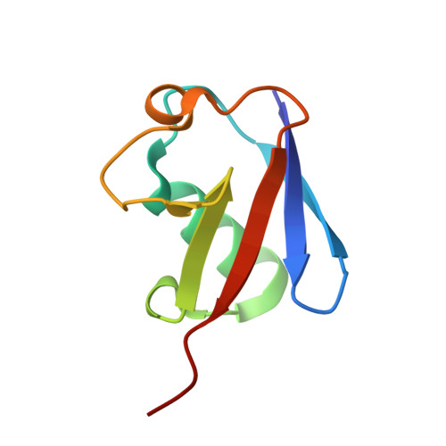
3H7S

Crystal structures of K63-linked di- and tri-ubiquitin reveal a highly extended chain architecture


Experimental Data Snapshot

  • Method: X-RAY DIFFRACTION
  • Resolution: 2.30 Å
  • R-Value Free: 0.253 
  • R-Value Work: 0.196 
  • R-Value Observed: 0.199 

wwPDB Validation   3D Report Full Report


This is version 1.3 of the entry. See complete history


Literature

Crystal structures of Lys-63-linked tri- and di-ubiquitin reveal a highly extended chain architecture.

Weeks, S.D.Grasty, K.C.Hernandez-Cuebas, L.Loll, P.J.

(2009) Proteins 77: 753-759

  • DOI: https://doi.org/10.1002/prot.22568
  • Primary Citation of Related Structures:  
    3H7P, 3H7S

  • PubMed Abstract: 

    The covalent attachment of different types of poly-ubiquitin chains signal different outcomes for the proteins so targeted. For example, a protein modified with Lys-48-linked poly-ubiquitin chains is targeted for proteasomal degradation, whereas Lys-63-linked chains encode nondegradative signals. The structural features that enable these different types of chains to encode different signals have not yet been fully elucidated. We report here the X-ray crystal structures of Lys-63-linked tri- and di-ubiquitin at resolutions of 2.3 and 1.9 A, respectively. The tri- and di-ubiquitin species adopt essentially identical structures. In both instances, the ubiquitin chain assumes a highly extended conformation with a left-handed helical twist; the helical chain contains four ubiquitin monomers per turn and has a repeat length of approximately 110 A. Interestingly, Lys-48 ubiquitin chains also adopt a left-handed helical structure with a similar repeat length. However, the Lys-63 architecture is much more open than that of Lys-48 chains and exposes much more of the ubiquitin surface for potential recognition events. These new crystal structures are consistent with the results of solution studies of Lys-63 chain conformation, and reveal the structural basis for differential recognition of Lys-63 versus Lys-48 chains.


  • Organizational Affiliation

    Department of Biochemistry and Molecular Biology, Drexel University College of Medicine, Philadelphia, Pennsylvania 19102, USA.


Macromolecules
Find similar proteins by:  (by identity cutoff)  |  3D Structure
Entity ID: 1
MoleculeChains Sequence LengthOrganismDetailsImage
Ubiquitin
A, B
76Homo sapiensMutation(s): 1 
Gene Names: RPS27AUBA52UBA80UBBUBCUBCEP1UBCEP2
UniProt & NIH Common Fund Data Resources
Find proteins for P0CG48 (Homo sapiens)
Explore P0CG48 
Go to UniProtKB:  P0CG48
PHAROS:  P0CG48
GTEx:  ENSG00000150991 
Entity Groups  
Sequence Clusters30% Identity50% Identity70% Identity90% Identity95% Identity100% Identity
UniProt GroupP0CG48
Sequence Annotations
Expand
  • Reference Sequence
Experimental Data & Validation

Experimental Data

  • Method: X-RAY DIFFRACTION
  • Resolution: 2.30 Å
  • R-Value Free: 0.253 
  • R-Value Work: 0.196 
  • R-Value Observed: 0.199 
  • Space Group: P 43 3 2
Unit Cell:
Length ( Å )Angle ( ˚ )
a = 105.32α = 90
b = 105.32β = 90
c = 105.32γ = 90
Software Package:
Software NamePurpose
XSCALEdata scaling
PHASERphasing
PHENIXrefinement
PDB_EXTRACTdata extraction
Blu-Icedata collection
XDSdata reduction

Structure Validation

View Full Validation Report


Entry History 

Deposition Data

Revision History  (Full details and data files)

  • Version 1.0: 2009-09-22
    Type: Initial release
  • Version 1.1: 2011-07-13
    Changes: Version format compliance
  • Version 1.2: 2017-11-01
    Changes: Advisory, Refinement description
  • Version 1.3: 2024-02-21
    Changes: Advisory, Data collection, Database references, Derived calculations