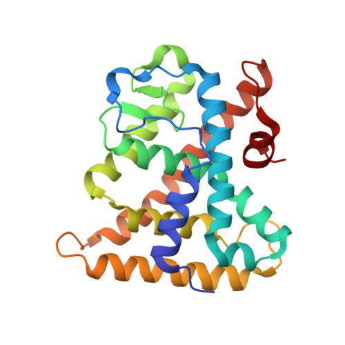
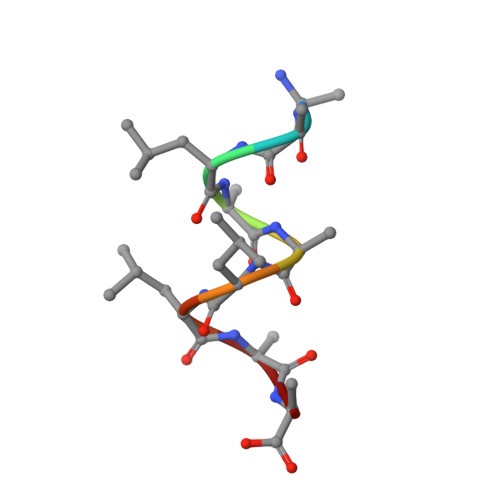
3FS1

Crystal structure of HNF4a LBD in complex with the ligand and the coactivator PGC-1a fragment


Experimental Data Snapshot

  • Method: X-RAY DIFFRACTION
  • Resolution: 2.20 Å
  • R-Value Free: 0.236 
  • R-Value Work: 0.190 
  • R-Value Observed: 0.195 

Starting Model: experimental
View more details

wwPDB Validation   3D Report Full Report


Ligand Structure Quality Assessment 


This is version 1.3 of the entry. See complete history


Literature

Multiple binding modes between HNF4alpha and the LXXLL motifs of PGC-1alpha lead to full activation

Rha, G.B.Wu, G.Shoelson, S.E.Chi, Y.I.

(2009) J Biol Chem 284: 35165-35176

  • DOI: https://doi.org/10.1074/jbc.M109.052506
  • Primary Citation of Related Structures:  
    3FS1

  • PubMed Abstract: 

    Hepatocyte nuclear factor 4alpha (HNF4alpha) is a novel nuclear receptor that participates in a hierarchical network of transcription factors regulating the development and physiology of such vital organs as the liver, pancreas, and kidney. Among the various transcriptional coregulators with which HNF4alpha interacts, peroxisome proliferation-activated receptor gamma (PPARgamma) coactivator 1alpha (PGC-1alpha) represents a novel coactivator whose activation is unusually robust and whose binding mode appears to be distinct from that of canonical coactivators such as NCoA/SRC/p160 family members. To elucidate the potentially unique molecular mechanism of PGC-1alpha recruitment, we have determined the crystal structure of HNF4alpha in complex with a fragment of PGC-1alpha containing all three of its LXXLL motifs. Despite the presence of all three LXXLL motifs available for interactions, only one is bound at the canonical binding site, with no additional contacts observed between the two proteins. However, a close inspection of the electron density map indicates that the bound LXXLL motif is not a selected one but an averaged structure of more than one LXXLL motif. Further biochemical and functional studies show that the individual LXXLL motifs can bind but drive only minimal transactivation. Only when more than one LXXLL motif is involved can significant transcriptional activity be measured, and full activation requires all three LXXLL motifs. These findings led us to propose a model wherein each LXXLL motif has an additive effect, and the multiple binding modes by HNF4alpha toward the LXXLL motifs of PGC-1alpha could account for the apparent robust activation by providing a flexible mechanism for combinatorial recruitment of additional coactivators and mediators.


  • Organizational Affiliation

    Department of Molecular and Cellular Biochemistry, Center for Structural Biology, University of Kentucky, Lexington, Kentucky 40536, USA.


Macromolecules
Find similar proteins by:  (by identity cutoff)  |  3D Structure
Entity ID: 1
MoleculeChains Sequence LengthOrganismDetailsImage
Hepatocyte nuclear factor 4-alpha230Homo sapiensMutation(s): 0 
Gene Names: HNF4HNF4ANR2A1TCF14
UniProt & NIH Common Fund Data Resources
Find proteins for P41235 (Homo sapiens)
Explore P41235 
Go to UniProtKB:  P41235
PHAROS:  P41235
GTEx:  ENSG00000101076 
Entity Groups  
Sequence Clusters30% Identity50% Identity70% Identity90% Identity95% Identity100% Identity
UniProt GroupP41235
Sequence Annotations
Expand
  • Reference Sequence

Find similar proteins by:  Sequence   |   3D Structure  

Entity ID: 2
MoleculeChains Sequence LengthOrganismDetailsImage
PPARgamma Coactivator-1a (PGC-1a)9Homo sapiensMutation(s): 0 
Gene Names: PGC-1a
Sequence Annotations
Expand
  • Reference Sequence
Small Molecules
Ligands 1 Unique
IDChains Name / Formula / InChI Key2D Diagram3D Interactions
MYR
Query on MYR

Download Ideal Coordinates CCD File 
C [auth A]MYRISTIC ACID
C14 H28 O2
TUNFSRHWOTWDNC-UHFFFAOYSA-N
Experimental Data & Validation

Experimental Data

  • Method: X-RAY DIFFRACTION
  • Resolution: 2.20 Å
  • R-Value Free: 0.236 
  • R-Value Work: 0.190 
  • R-Value Observed: 0.195 
  • Space Group: P 42 2 2
Unit Cell:
Length ( Å )Angle ( ˚ )
a = 113.049α = 90
b = 113.049β = 90
c = 57.322γ = 90
Software Package:
Software NamePurpose
DENZOdata reduction
SCALEPACKdata scaling
MOLREPphasing
REFMACrefinement
PDB_EXTRACTdata extraction
HKL-2000data collection

Structure Validation

View Full Validation Report


Ligand Structure Quality Assessment 


Entry History 

Deposition Data

Revision History  (Full details and data files)

  • Version 1.0: 2009-10-27
    Type: Initial release
  • Version 1.1: 2011-07-13
    Changes: Version format compliance
  • Version 1.2: 2017-11-01
    Changes: Refinement description
  • Version 1.3: 2023-09-06
    Changes: Data collection, Database references, Derived calculations, Refinement description