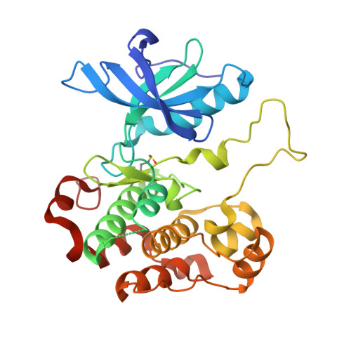
3D94

Crystal structure of the insulin-like growth factor-1 receptor kinase in complex with PQIP


Experimental Data Snapshot

  • Method: X-RAY DIFFRACTION
  • Resolution: 2.30 Å
  • R-Value Free: 0.236 
  • R-Value Work: 0.198 
  • R-Value Observed: 0.200 

Starting Model: experimental
View more details

wwPDB Validation   3D Report Full Report


Ligand Structure Quality Assessment 


This is version 1.3 of the entry. See complete history


Literature

Small-molecule inhibition and activation-loop trans-phosphorylation of the IGF1 receptor

Wu, J.Li, W.Craddock, B.P.Foreman, K.W.Mulvihill, M.J.Ji, Q.S.Miller, W.T.Hubbard, S.R.

(2008) EMBO J 27: 1985-1994

  • DOI: https://doi.org/10.1038/emboj.2008.116
  • Primary Citation of Related Structures:  
    3D94

  • PubMed Abstract: 

    The insulin-like growth factor-1 receptor (IGF1R) is a receptor tyrosine kinase (RTK) that has a critical role in mitogenic signalling during embryogenesis and an antiapoptotic role in the survival and progression of many human tumours. Here, we present the crystal structure of the tyrosine kinase domain of IGF1R (IGF1RK), in its unphosphorylated state, in complex with a novel compound, cis-3-[3-(4-methyl-piperazin-l-yl)-cyclobutyl]-1-(2-phenyl-quinolin-7-yl)-imidazo[1,5-a]pyrazin-8-ylamine (PQIP), which we show is a potent inhibitor of both the unphosphorylated (basal) and phosphorylated (activated) states of the kinase. PQIP interacts with residues in the ATP-binding pocket and in the activation loop, which confers specificity for IGF1RK and the highly related insulin receptor (IR) kinase. In this crystal structure, the IGF1RK active site is occupied by Tyr1135 from the activation loop of an symmetry (two-fold)-related molecule. This dimeric arrangement affords, for the first time, a visualization of the initial trans-phosphorylation event in the activation loop of an RTK, and provides a molecular rationale for a naturally occurring mutation in the activation loop of the IR that causes type II diabetes mellitus.


  • Organizational Affiliation

    Structural Biology Program, Kimmel Center for Biology and Medicine of the Skirball Institute of Biomolecular Medicine, Department of Pharmacology, New York University School of Medicine, New York, NY 10016, USA.


Macromolecules
Find similar proteins by:  (by identity cutoff)  |  3D Structure
Entity ID: 1
MoleculeChains Sequence LengthOrganismDetailsImage
Insulin-like growth factor 1 receptor beta chain301Homo sapiensMutation(s): 1 
Gene Names: IGF1R
EC: 2.7.1.112 (PDB Primary Data), 2.7.10.1 (PDB Primary Data)
UniProt & NIH Common Fund Data Resources
Find proteins for P08069 (Homo sapiens)
Explore P08069 
Go to UniProtKB:  P08069
PHAROS:  P08069
GTEx:  ENSG00000140443 
Entity Groups  
Sequence Clusters30% Identity50% Identity70% Identity90% Identity95% Identity100% Identity
UniProt GroupP08069
Sequence Annotations
Expand
  • Reference Sequence
Small Molecules
Ligands 2 Unique
IDChains Name / Formula / InChI Key2D Diagram3D Interactions
D94
Query on D94

Download Ideal Coordinates CCD File 
C [auth A]3-[cis-3-(4-methylpiperazin-1-yl)cyclobutyl]-1-(2-phenylquinolin-7-yl)imidazo[1,5-a]pyrazin-8-amine
C30 H31 N7
PDJARQSWGDDFHH-PSWAGMNNSA-N
CA
Query on CA

Download Ideal Coordinates CCD File 
B [auth A]CALCIUM ION
Ca
BHPQYMZQTOCNFJ-UHFFFAOYSA-N
Modified Residues  1 Unique
IDChains TypeFormula2D DiagramParent
MHO
Query on MHO
A
L-PEPTIDE LINKINGC5 H11 N O3 SMET
Binding Affinity Annotations 
IDSourceBinding Affinity
D94 PDBBind:  3D94 Ki: 157 (nM) from 1 assay(s)
BindingDB:  3D94 IC50: min: 19, max: 65 (nM) from 4 assay(s)
Experimental Data & Validation

Experimental Data

  • Method: X-RAY DIFFRACTION
  • Resolution: 2.30 Å
  • R-Value Free: 0.236 
  • R-Value Work: 0.198 
  • R-Value Observed: 0.200 
  • Space Group: H 3 2
Unit Cell:
Length ( Å )Angle ( ˚ )
a = 208.182α = 90
b = 208.182β = 90
c = 50.853γ = 120
Software Package:
Software NamePurpose
REFMACrefinement
ADSCdata collection
HKL-2000data reduction
HKL-2000data scaling
AMoREphasing

Structure Validation

View Full Validation Report


Ligand Structure Quality Assessment 


Entry History 

Deposition Data

Revision History  (Full details and data files)

  • Version 1.0: 2008-07-29
    Type: Initial release
  • Version 1.1: 2011-07-13
    Changes: Version format compliance
  • Version 1.2: 2023-08-30
    Changes: Data collection, Database references, Derived calculations, Refinement description
  • Version 1.3: 2024-10-30
    Changes: Structure summary