3D5S

Crystal Structure of Efb-C (R131A) / C3d Complex


Experimental Data Snapshot

  • Method: X-RAY DIFFRACTION
  • Resolution: 2.30 Å
  • R-Value Free: 0.218 
  • R-Value Work: 0.204 

Starting Model: experimental
View more details

wwPDB Validation   3D Report Full Report


This is version 1.4 of the entry. See complete history


Literature

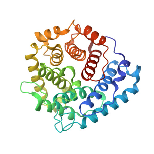
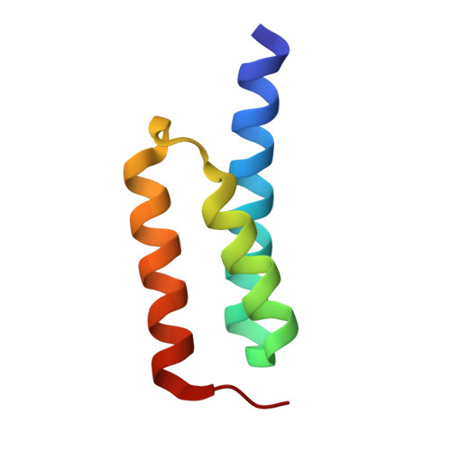
Electrostatic contributions drive the interaction between Staphylococcus aureus protein Efb-C and its complement target C3d.

Haspel, N.Ricklin, D.Geisbrecht, B.V.Kavraki, L.E.Lambris, J.D.

(2008) Protein Sci 17: 1894-1906

  • DOI: https://doi.org/10.1110/ps.036624.108
  • Primary Citation of Related Structures:  
    3D5R, 3D5S

  • PubMed Abstract: 

    The C3-inhibitory domain of Staphylococcus aureus extracellular fibrinogen-binding protein (Efb-C) defines a novel three-helix bundle motif that regulates complement activation. Previous crystallographic studies of Efb-C bound to its cognate subdomain of human C3 (C3d) identified Arg-131 and Asn-138 of Efb-C as key residues for its activity. In order to characterize more completely the physical and chemical driving forces behind this important interaction, we employed in this study a combination of structural, biophysical, and computational methods to analyze the interaction of C3d with Efb-C and the single-point mutants R131A and N138A. Our results show that while these mutations do not drastically affect the structure of the Efb-C/C3d recognition complex, they have significant adverse effects on both the thermodynamic and kinetic profiles of the resulting complexes. We also characterized other key interactions along the Efb-C/C3d binding interface and found an intricate network of salt bridges and hydrogen bonds that anchor Efb-C to C3d, resulting in its potent complement inhibitory properties.


  • Organizational Affiliation

    Department of Computer Science, Rice University, Houston, Texas 77005, USA.


Macromolecules
Find similar proteins by:  (by identity cutoff)  |  3D Structure
Entity ID: 1
MoleculeChains Sequence LengthOrganismDetailsImage
Complement C3A,
C [auth B]
297Homo sapiensMutation(s): 1 
Gene Names: C3CPAMD1
UniProt & NIH Common Fund Data Resources
Find proteins for P01024 (Homo sapiens)
Explore P01024 
Go to UniProtKB:  P01024
GTEx:  ENSG00000125730 
Entity Groups  
Sequence Clusters30% Identity50% Identity70% Identity90% Identity95% Identity100% Identity
UniProt GroupP01024
Sequence Annotations
Expand
  • Reference Sequence
Find similar proteins by:  (by identity cutoff)  |  3D Structure
Entity ID: 2
MoleculeChains Sequence LengthOrganismDetailsImage
Fibrinogen-binding proteinB [auth C],
D
65Staphylococcus aureus subsp. aureus str. NewmanMutation(s): 1 
Gene Names: fibefbNWMN_1069
UniProt
Find proteins for A6QG59 (Staphylococcus aureus (strain Newman))
Explore A6QG59 
Go to UniProtKB:  A6QG59
Entity Groups  
Sequence Clusters30% Identity50% Identity70% Identity90% Identity95% Identity100% Identity
UniProt GroupA6QG59
Sequence Annotations
Expand
  • Reference Sequence
Experimental Data & Validation

Experimental Data

  • Method: X-RAY DIFFRACTION
  • Resolution: 2.30 Å
  • R-Value Free: 0.218 
  • R-Value Work: 0.204 
  • Space Group: P 41
Unit Cell:
Length ( Å )Angle ( ˚ )
a = 90.89α = 90
b = 90.89β = 90
c = 122.83γ = 90
Software Package:
Software NamePurpose
DENZOdata reduction
SCALEPACKdata scaling
MOLREPphasing
CNSrefinement
PDB_EXTRACTdata extraction
SERGUIdata collection
HKL-2000data scaling

Structure Validation

View Full Validation Report


Entry History 

Deposition Data

Revision History  (Full details and data files)

  • Version 1.0: 2008-09-16
    Type: Initial release
  • Version 1.1: 2011-07-13
    Changes: Version format compliance
  • Version 1.2: 2017-10-25
    Changes: Refinement description
  • Version 1.3: 2021-10-20
    Changes: Database references
  • Version 1.4: 2023-08-30
    Changes: Data collection, Refinement description