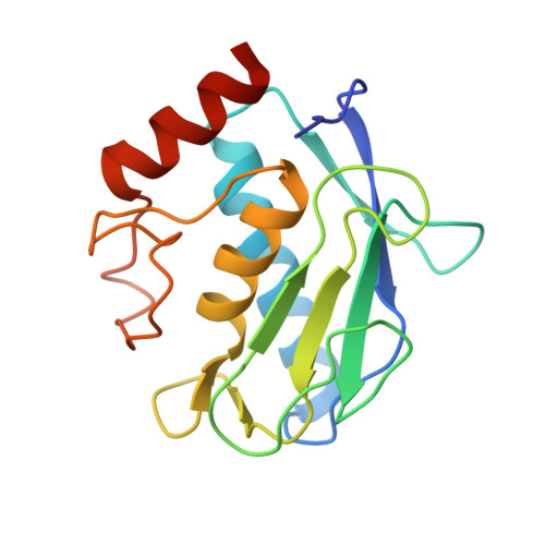
2W0D

Does a Fast Nuclear Magnetic Resonance Spectroscopy- and X-Ray Crystallography Hybrid Approach Provide Reliable Structural Information of Ligand-Protein Complexes? A Case Study of Metalloproteinases.


Experimental Data Snapshot

  • Method: X-RAY DIFFRACTION
  • Resolution: 2.00 Å
  • R-Value Free: 0.237 
  • R-Value Work: 0.179 
  • R-Value Observed: 0.182 

Starting Model: other
View more details

wwPDB Validation   3D Report Full Report


Ligand Structure Quality Assessment 


This is version 1.4 of the entry. See complete history


Literature

Does a Fast Nuclear Magnetic Resonance Spectroscopy- and X-Ray Crystallography Hybrid Approach Provide Reliable Structural Information of Ligand-Protein Complexes? a Case Study of Metalloproteinases.

Isaksson, J.Nystrom, S.Derbyshire, D.J.Wallberg, H.Agback, T.Kovacs, H.Bertini, I.Felli, I.C.

(2009) J Med Chem 52: 1712

  • DOI: https://doi.org/10.1021/jm801388q
  • Primary Citation of Related Structures:  
    2W0D

  • PubMed Abstract: 

    A human matrix metalloproteinase (MMP) hydroxamic acid inhibitor (CGS27023A) was cross-docked into 15 MMP-12, MMP-13, MMP-9, and MMP-1 cocrystal structures. The aim was to validate a fast protocol for ligand binding conformation elucidation and to probe the feasibility of using inhibitor-protein NMR contacts to dock an inhibitor into related MMP crystal structures. Such an approach avoids full NMR structure elucidation, saving both spectrometer- and analysis time. We report here that for the studied MMPs, one can obtain docking results well within 1 A compared to the corresponding reference X-ray structure, using backbone amide contacts only. From the perspective of the pharmaceutical industry, these results are relevant for the binding studies of inhibitor series to a common target and have the potential advantage of obtaining information on protein-inhibitor complexes that are difficult to crystallize.


  • Organizational Affiliation

    Medivir AB, PO Box 1086, SE-141 22 Huddinge, Sweden. johan.isaksson@medivir.se


Macromolecules
Find similar proteins by:  (by identity cutoff)  |  3D Structure
Entity ID: 1
MoleculeChains Sequence LengthOrganismDetailsImage
MACROPHAGE METALLOELASTASE
A, B, C, D
164Homo sapiensMutation(s): 1 
EC: 3.4.24.65
UniProt & NIH Common Fund Data Resources
Find proteins for P39900 (Homo sapiens)
Explore P39900 
Go to UniProtKB:  P39900
PHAROS:  P39900
GTEx:  ENSG00000262406 
Entity Groups  
Sequence Clusters30% Identity50% Identity70% Identity90% Identity95% Identity100% Identity
UniProt GroupP39900
Sequence Annotations
Expand
  • Reference Sequence
Small Molecules
Ligands 6 Unique
IDChains Name / Formula / InChI Key2D Diagram3D Interactions
CGS
Query on CGS

Download Ideal Coordinates CCD File 
DA [auth C],
N [auth A],
NA [auth D],
T [auth B]
N-HYDROXY-2(R)-[[(4-METHOXYPHENYL)SULFONYL](3-PICOLYL)AMINO]-3-METHYLBUTANAMIDE HYDROCHLORIDE
C18 H23 N3 O5 S
BSIZUMJRKYHEBR-QGZVFWFLSA-N
ZN
Query on ZN

Download Ideal Coordinates CCD File 
E [auth A]
EA [auth D]
F [auth A]
FA [auth D]
J [auth A]
E [auth A],
EA [auth D],
F [auth A],
FA [auth D],
J [auth A],
O [auth B],
P [auth B],
U [auth C],
V [auth C],
Z [auth C]
ZINC ION
Zn
PTFCDOFLOPIGGS-UHFFFAOYSA-N
ACT
Query on ACT

Download Ideal Coordinates CCD File 
AA [auth C]
CA [auth C]
K [auth A]
L [auth A]
LA [auth D]
AA [auth C],
CA [auth C],
K [auth A],
L [auth A],
LA [auth D],
M [auth A],
MA [auth D]
ACETATE ION
C2 H3 O2
QTBSBXVTEAMEQO-UHFFFAOYSA-M
CA
Query on CA

Download Ideal Coordinates CCD File 
G [auth A]
GA [auth D]
H [auth A]
HA [auth D]
I [auth A]
G [auth A],
GA [auth D],
H [auth A],
HA [auth D],
I [auth A],
IA [auth D],
Q [auth B],
R [auth B],
S [auth B],
W [auth C],
X [auth C],
Y [auth C]
CALCIUM ION
Ca
BHPQYMZQTOCNFJ-UHFFFAOYSA-N
CL
Query on CL

Download Ideal Coordinates CCD File 
BA [auth C],
JA [auth D]
CHLORIDE ION
Cl
VEXZGXHMUGYJMC-UHFFFAOYSA-M
NA
Query on NA

Download Ideal Coordinates CCD File 
KA [auth D]SODIUM ION
Na
FKNQFGJONOIPTF-UHFFFAOYSA-N
Binding Affinity Annotations 
IDSourceBinding Affinity
CGS PDBBind:  2W0D IC50: 2 (nM) from 1 assay(s)
Experimental Data & Validation

Experimental Data

  • Method: X-RAY DIFFRACTION
  • Resolution: 2.00 Å
  • R-Value Free: 0.237 
  • R-Value Work: 0.179 
  • R-Value Observed: 0.182 
  • Space Group: P 1 21 1
Unit Cell:
Length ( Å )Angle ( ˚ )
a = 38.16α = 90
b = 99.939β = 96.59
c = 79.423γ = 90
Software Package:
Software NamePurpose
REFMACrefinement
MOSFLMdata reduction
SCALAdata scaling
PHASERphasing

Structure Validation

View Full Validation Report


Ligand Structure Quality Assessment 


Entry History 

Deposition Data

Revision History  (Full details and data files)

  • Version 1.0: 2009-03-03
    Type: Initial release
  • Version 1.1: 2011-07-13
    Changes: Refinement description, Version format compliance
  • Version 1.2: 2011-12-28
    Changes: Other
  • Version 1.3: 2019-05-22
    Changes: Data collection, Experimental preparation, Other
  • Version 1.4: 2024-05-01
    Changes: Data collection, Database references, Derived calculations, Other, Refinement description