2VX0

ephB4 kinase domain inhibitor complex


Experimental Data Snapshot

  • Method: X-RAY DIFFRACTION
  • Resolution: 2.10 Å
  • R-Value Free: 0.233 
  • R-Value Work: 0.184 
  • R-Value Observed: 0.187 

Starting Model: experimental
View more details

wwPDB Validation   3D Report Full Report


Ligand Structure Quality Assessment 


This is version 1.6 of the entry. See complete history


Literature

Inhibitors of the Tyrosine Kinase Ephb4. Part 1: Structure-Based Design and Optimization of a Series of 2,4-Bis-Anilinopyrimidines.

Bardelle, C.Cross, D.Davenport, S.Kettle, J.G.Ko, E.J.Leach, A.G.Mortlock, A.Read, J.Roberts, N.J.Robins, P.Williams, E.J.

(2008) Bioorg Med Chem Lett 18: 2776

  • DOI: https://doi.org/10.1016/j.bmcl.2008.04.015
  • Primary Citation of Related Structures:  
    2VWU, 2VWV, 2VWW, 2VX0

  • PubMed Abstract: 

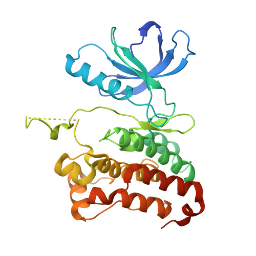
    A series of bis-anilinopyrimidines have been identified as potent inhibitors of the tyrosine kinase EphB4. Structural information from two alternative series identified from screening efforts was combined to identify the initial leads.


  • Organizational Affiliation

    AstraZeneca, Mereside, Alderley Park, Macclesfield, Cheshire SK10 4TG, UK.


Macromolecules
Find similar proteins by:  (by identity cutoff)  |  3D Structure
Entity ID: 1
MoleculeChains Sequence LengthOrganismDetailsImage
EPHRIN TYPE-B RECEPTOR 4302Homo sapiensMutation(s): 1 
EC: 2.7.10.1
UniProt & NIH Common Fund Data Resources
Find proteins for P54760 (Homo sapiens)
Explore P54760 
Go to UniProtKB:  P54760
GTEx:  ENSG00000196411 
Entity Groups  
Sequence Clusters30% Identity50% Identity70% Identity90% Identity95% Identity100% Identity
UniProt GroupP54760
Sequence Annotations
Expand
  • Reference Sequence
Small Molecules
Binding Affinity Annotations 
IDSourceBinding Affinity
7X7 PDBBind:  2VX0 IC50: 580 (nM) from 1 assay(s)
BindingDB:  2VX0 IC50: 580 (nM) from 1 assay(s)
Experimental Data & Validation

Experimental Data

  • Method: X-RAY DIFFRACTION
  • Resolution: 2.10 Å
  • R-Value Free: 0.233 
  • R-Value Work: 0.184 
  • R-Value Observed: 0.187 
  • Space Group: P 1 21 1
Unit Cell:
Length ( Å )Angle ( ˚ )
a = 46.123α = 90
b = 53.492β = 110.91
c = 61.265γ = 90
Software Package:
Software NamePurpose
REFMACrefinement
CrystalCleardata reduction
CrystalCleardata scaling
AMoREphasing

Structure Validation

View Full Validation Report


Ligand Structure Quality Assessment 


Entry History 

Deposition Data

Revision History  (Full details and data files)

  • Version 1.0: 2008-10-28
    Type: Initial release
  • Version 1.1: 2011-05-08
    Changes: Version format compliance
  • Version 1.2: 2011-07-13
    Changes: Version format compliance
  • Version 1.3: 2017-06-28
    Changes: Data collection
  • Version 1.4: 2019-04-24
    Changes: Data collection, Source and taxonomy
  • Version 1.5: 2019-05-08
    Changes: Data collection, Experimental preparation
  • Version 1.6: 2023-12-13
    Changes: Data collection, Database references, Derived calculations, Other, Refinement description