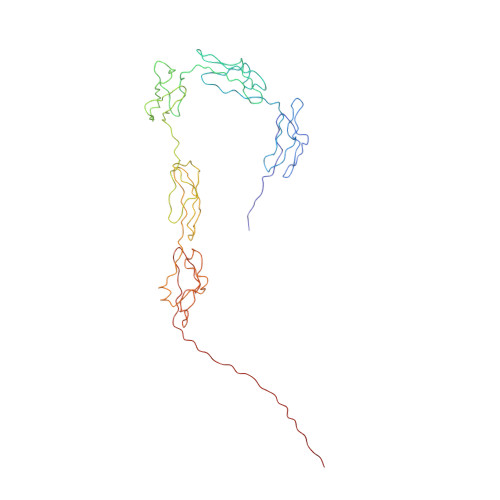
The regulatory SCR-1/5 and cell surface-binding SCR-16/20 fragments of factor H reveal partially folded-back solution structures and different self-associative properties.
Okemefuna, A.I., Gilbert, H.E., Griggs, K.M., Ormsby, R.J., Gordon, D.L., Perkins, S.J.(2008) J Mol Biol 375: 80-101
- PubMed: 18005991
- DOI: https://doi.org/10.1016/j.jmb.2007.09.026
- Primary Citation of Related Structures:
2QFG, 2QFH - PubMed Abstract:
Factor H (FH) is a plasma glycoprotein that plays a central role in regulation of the alternative pathway of complement. It is composed of 20 short complement regulator (SCR) domains. The SCR-1/5 fragment is required for decay acceleration and cofactor activity, while the SCR-16/20 fragment possesses binding sites for complement C3d and heparin. X-ray scattering and analytical ultracentrifugation showed that SCR-1/5 was monomeric, while SCR-16/20 formed dimers. The Guinier radius of gyration R(G) of 4.3 nm for SCR-1/5 and those of 4.7 nm and about 7.8 nm for monomeric and dimeric SCR-16/20, respectively, showed that their structures are partially folded back and bent. The distance distribution function P(r) showed that SCR-1/5 has a maximum dimension of 15 nm while monomeric and dimeric SCR-16/20 are 17 nm and about 27 nm long, respectively. The sedimentation coefficient of 2.4 S for SCR-1/5 showed no concentration-dependence, while that for SCR-16/20 was 2.8 S for the monomer and 3.9 S for the dimer. Sedimentation equilibrium data showed that SCR-1/5 is monomeric while SCR-16/20 exhibited a weak monomer-dimer equilibrium with a dissociation constant of 16 microM. The constrained scattering and sedimentation modelling of SCR-1/5 and SCR-16/20 showed that partially folded-back and bent flexible SCR arrangements fitted both data sets better than extended linear arrangements, and that the dimer was best modelled in the SCR-16/20 model by an end-to-end association of two SCR-20 domains. The SCR-1/5 and SCR-16/20 models were conformationally similar to the previously determined partially folded-back structure for intact wild-type FH, hence suggesting a partial explanation of the intact FH structure. Comparison of the SCR-16/20 model with the crystal structure of C3b clarified reasons for the distribution of mutations leading to atypical haemolytic uraemic syndrome.
Organizational Affiliation:
Department of Biochemistry and Molecular Biology, Darwin Building, University College London, Gower Street, London WC1E 6BT, UK.


