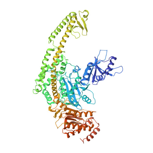
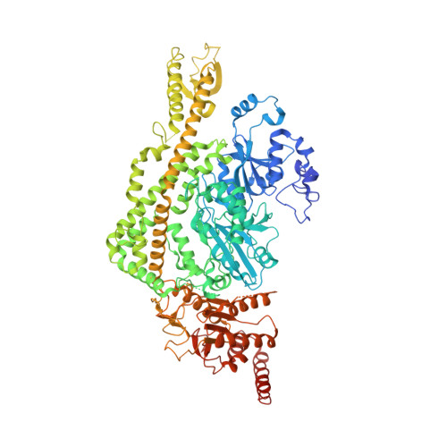
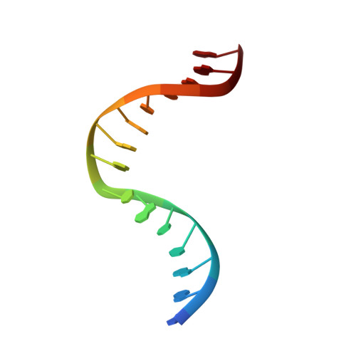
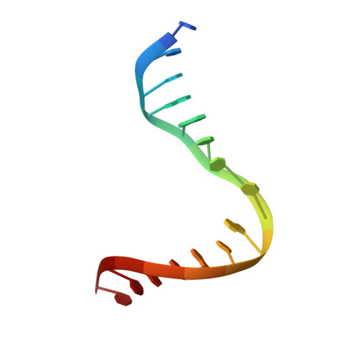
2O8E

human MutSalpha (MSH2/MSH6) bound to a G T mispair, with ADP bound to MSH2 only


Experimental Data Snapshot

  • Method: X-RAY DIFFRACTION
  • Resolution: 3.30 Å
  • R-Value Free: 0.291 
  • R-Value Work: 0.253 
  • R-Value Observed: 0.255 

Starting Model: experimental
View more details

wwPDB Validation   3D Report Full Report


Ligand Structure Quality Assessment 


This is version 1.4 of the entry. See complete history


Literature

Structure of the Human MutSalpha DNA Lesion Recognition Complex.

Warren, J.J.Pohlhaus, T.J.Changela, A.Iyer, R.R.Modrich, P.L.Beese, L.S.

(2007) Mol Cell 26: 579-592

  • DOI: https://doi.org/10.1016/j.molcel.2007.04.018
  • Primary Citation of Related Structures:  
    2O8B, 2O8C, 2O8D, 2O8E, 2O8F

  • PubMed Abstract: 

    Mismatch repair (MMR) ensures the fidelity of DNA replication, initiates the cellular response to certain classes of DNA damage, and has been implicated in the generation of immune diversity. Each of these functions depends on MutSalpha (MSH2*MSH6 heterodimer). Inactivation of this protein complex is responsible for tumor development in about half of known hereditary nonpolyposis colorectal cancer kindreds and also occurs in sporadic tumors in a variety of tissues. Here, we describe a series of crystal structures of human MutSalpha bound to different DNA substrates, each known to elicit one of the diverse biological responses of the MMR pathway. All lesions are recognized in a similar manner, indicating that diversity of MutSalpha-dependent responses to DNA lesions is generated in events downstream of this lesion recognition step. This study also allows rigorous mapping of cancer-causing mutations and furthermore suggests structural pathways for allosteric communication between different regions within the heterodimer.


  • Organizational Affiliation

    Department of Biochemistry, Duke University Medical Center, Durham, NC 27710, USA.


Macromolecules

Find similar proteins by:  (by identity cutoff)  |  3D Structure
Entity ID: 3
MoleculeChains Sequence LengthOrganismDetailsImage
DNA mismatch repair protein Msh2C [auth A]934Homo sapiensMutation(s): 1 
Gene Names: MSH2
UniProt & NIH Common Fund Data Resources
Find proteins for P43246 (Homo sapiens)
Explore P43246 
Go to UniProtKB:  P43246
PHAROS:  P43246
GTEx:  ENSG00000095002 
Entity Groups  
Sequence Clusters30% Identity50% Identity70% Identity90% Identity95% Identity100% Identity
UniProt GroupP43246
Sequence Annotations
Expand
  • Reference Sequence
Find similar proteins by:  (by identity cutoff)  |  3D Structure
Entity ID: 4
MoleculeChains Sequence LengthOrganismDetailsImage
DNA mismatch repair protein MSH6D [auth B]1,022Homo sapiensMutation(s): 0 
Gene Names: MSH6GTBP
UniProt & NIH Common Fund Data Resources
Find proteins for P52701 (Homo sapiens)
Explore P52701 
Go to UniProtKB:  P52701
PHAROS:  P52701
GTEx:  ENSG00000116062 
Entity Groups  
Sequence Clusters30% Identity50% Identity70% Identity90% Identity95% Identity100% Identity
UniProt GroupP52701
Sequence Annotations
Expand
  • Reference Sequence

Find similar nucleic acids by:  Sequence   |   3D Structure  

Entity ID: 1
MoleculeChains LengthOrganismImage
5'-D(*GP*AP*AP*CP*CP*GP*CP*GP*GP*GP*CP*TP*AP*GP*G)-3'A [auth E]15N/A
Sequence Annotations
Expand
  • Reference Sequence

Find similar nucleic acids by:  Sequence   |   3D Structure  

Entity ID: 2
MoleculeChains LengthOrganismImage
5'-D(*CP*CP*TP*AP*GP*CP*CP*TP*GP*CP*GP*GP*TP*TP*C)-3'B [auth F]15N/A
Sequence Annotations
Expand
  • Reference Sequence
Small Molecules
Ligands 2 Unique
IDChains Name / Formula / InChI Key2D Diagram3D Interactions
ADP
Query on ADP

Download Ideal Coordinates CCD File 
F [auth A]ADENOSINE-5'-DIPHOSPHATE
C10 H15 N5 O10 P2
XTWYTFMLZFPYCI-KQYNXXCUSA-N
MG
Query on MG

Download Ideal Coordinates CCD File 
E [auth A]MAGNESIUM ION
Mg
JLVVSXFLKOJNIY-UHFFFAOYSA-N
Experimental Data & Validation

Experimental Data

  • Method: X-RAY DIFFRACTION
  • Resolution: 3.30 Å
  • R-Value Free: 0.291 
  • R-Value Work: 0.253 
  • R-Value Observed: 0.255 
  • Space Group: P 43 3 2
Unit Cell:
Length ( Å )Angle ( ˚ )
a = 257.51α = 90
b = 257.51β = 90
c = 257.51γ = 90
Software Package:
Software NamePurpose
SCALAdata scaling
REFMACrefinement
PDB_EXTRACTdata extraction
MAR345dtbdata collection
XDSdata reduction
REFMACphasing

Structure Validation

View Full Validation Report


Ligand Structure Quality Assessment 


Entry History 

Deposition Data

Revision History  (Full details and data files)

  • Version 1.0: 2007-06-05
    Type: Initial release
  • Version 1.1: 2008-05-01
    Changes: Version format compliance
  • Version 1.2: 2011-07-13
    Changes: Advisory, Refinement description, Version format compliance
  • Version 1.3: 2021-10-20
    Changes: Database references, Derived calculations
  • Version 1.4: 2023-08-30
    Changes: Data collection, Refinement description