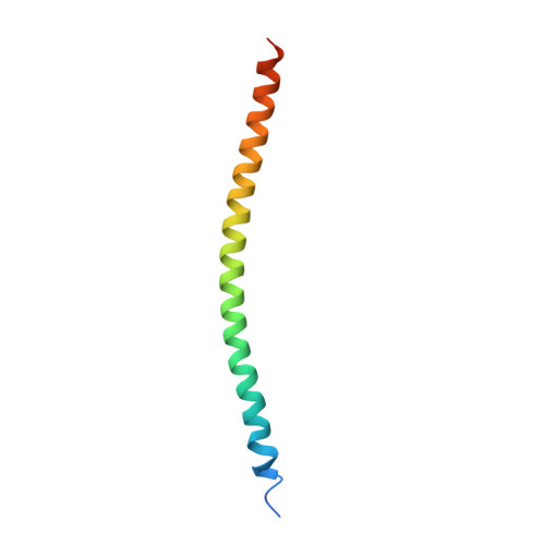
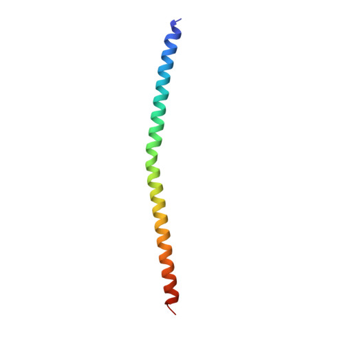
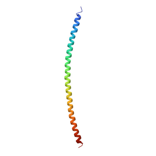
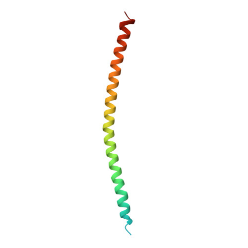
2NPS

Crystal Structure of the Early Endosomal SNARE Complex


Experimental Data Snapshot

  • Method: X-RAY DIFFRACTION
  • Resolution: 2.50 Å
  • R-Value Free: 0.289 
  • R-Value Work: 0.247 
  • R-Value Observed: 0.251 

Starting Model: experimental
View more details

wwPDB Validation   3D Report Full Report


This is version 1.4 of the entry. See complete history


Literature

Early endosomal SNAREs form a structurally conserved SNARE complex and fuse liposomes with multiple topologies

Zwilling, D.Cypionka, A.Pohl, W.H.Fasshauer, D.Walla, P.J.Wahl, M.C.Jahn, R.

(2007) EMBO J 26: 9-18

  • DOI: https://doi.org/10.1038/sj.emboj.7601467
  • Primary Citation of Related Structures:  
    2NPS

  • PubMed Abstract: 

    SNARE proteins mediate membrane fusion in eukaryotic cells. They contain conserved SNARE motifs that are usually located adjacent to a C-terminal transmembrane domain. SNARE motifs spontaneously assemble into four helix bundles, with each helix belonging to a different subfamily. Liposomes containing SNAREs spontaneously fuse with each other, but it is debated how the SNAREs are distributed between the membranes. Here, we report that the SNAREs mediating homotypic fusion of early endosomes fuse liposomes in five out of seven possible combinations, in contrast to previously studied SNAREs involved in heterotypic fusion events. The crystal structure of the early endosomal SNARE complex resembles that of the neuronal and late endosomal complexes, but differs in surface side-chain interactions. We conclude that homotypic fusion reactions may proceed with multiple SNARE topologies, suggesting that the conserved SNARE structure allows for flexibility in the initial interactions needed for fusion.


  • Organizational Affiliation

    Department of Neurobiology, Max-Planck Institute for Biophysical Chemistry, Göttingen, Germany.


Macromolecules
Find similar proteins by:  (by identity cutoff)  |  3D Structure
Entity ID: 1
MoleculeChains Sequence LengthOrganismDetailsImage
Vesicle-associated membrane protein 474Mus musculusMutation(s): 0 
Gene Names: Vamp4_predicted
Membrane Entity: Yes 
UniProt & NIH Common Fund Data Resources
Find proteins for O70480 (Mus musculus)
Explore O70480 
Go to UniProtKB:  O70480
IMPC:  MGI:1858730
Entity Groups  
Sequence Clusters30% Identity50% Identity70% Identity90% Identity95% Identity100% Identity
UniProt GroupO70480
Sequence Annotations
Expand
  • Reference Sequence
Find similar proteins by:  (by identity cutoff)  |  3D Structure
Entity ID: 2
MoleculeChains Sequence LengthOrganismDetailsImage
Syntaxin 1371Rattus norvegicusMutation(s): 0 
Gene Names: syntaxin 13
Membrane Entity: Yes 
UniProt
Find proteins for G3V7P1 (Rattus norvegicus)
Explore G3V7P1 
Go to UniProtKB:  G3V7P1
Entity Groups  
Sequence Clusters30% Identity50% Identity70% Identity90% Identity95% Identity100% Identity
UniProt GroupG3V7P1
Sequence Annotations
Expand
  • Reference Sequence
Find similar proteins by:  (by identity cutoff)  |  3D Structure
Entity ID: 3
MoleculeChains Sequence LengthOrganismDetailsImage
Vesicle transport through interaction with t-SNAREs homolog 1A81Rattus norvegicusMutation(s): 0 
Gene Names: Vti1a
Membrane Entity: Yes 
UniProt
Find proteins for Q9JI51 (Rattus norvegicus)
Explore Q9JI51 
Go to UniProtKB:  Q9JI51
Entity Groups  
Sequence Clusters30% Identity50% Identity70% Identity90% Identity95% Identity100% Identity
UniProt GroupQ9JI51
Sequence Annotations
Expand
  • Reference Sequence
Find similar proteins by:  (by identity cutoff)  |  3D Structure
Entity ID: 4
MoleculeChains Sequence LengthOrganismDetailsImage
Syntaxin-682Homo sapiensMutation(s): 0 
Gene Names: STX6
Membrane Entity: Yes 
UniProt & NIH Common Fund Data Resources
Find proteins for O43752 (Homo sapiens)
Explore O43752 
Go to UniProtKB:  O43752
PHAROS:  O43752
GTEx:  ENSG00000135823 
Entity Groups  
Sequence Clusters30% Identity50% Identity70% Identity90% Identity95% Identity100% Identity
UniProt GroupO43752
Sequence Annotations
Expand
  • Reference Sequence
Experimental Data & Validation

Experimental Data

  • Method: X-RAY DIFFRACTION
  • Resolution: 2.50 Å
  • R-Value Free: 0.289 
  • R-Value Work: 0.247 
  • R-Value Observed: 0.251 
  • Space Group: C 1 2 1
Unit Cell:
Length ( Å )Angle ( ˚ )
a = 252.882α = 90
b = 28.657β = 98.25
c = 41.887γ = 90
Software Package:
Software NamePurpose
MAR345dtbdata collection
MOLREPphasing
CNSrefinement
MAR345data collection
DENZOdata reduction
SCALEPACKdata scaling

Structure Validation

View Full Validation Report


Entry History 

Deposition Data

Revision History  (Full details and data files)

  • Version 1.0: 2006-12-26
    Type: Initial release
  • Version 1.1: 2008-05-01
    Changes: Version format compliance
  • Version 1.2: 2011-07-13
    Changes: Version format compliance
  • Version 1.3: 2017-10-18
    Changes: Advisory, Refinement description
  • Version 1.4: 2023-10-25
    Changes: Advisory, Data collection, Database references, Refinement description