2MSD

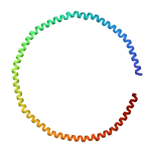
NMR data-driven model of GTPase KRas-GNP tethered to a lipid-bilayer nanodisc


Experimental Data Snapshot

  • Method: SOLUTION NMR
  • Conformers Calculated: 3000 
  • Conformers Submitted: 10 
  • Selection Criteria: structures with the lowest energy 

wwPDB Validation   3D Report Full Report


This is version 1.3 of the entry. See complete history


Literature

Oncogenic and RASopathy-associated K-RAS mutations relieve membrane-dependent occlusion of the effector-binding site.

Mazhab-Jafari, M.T.Marshall, C.B.Smith, M.J.Gasmi-Seabrook, G.M.Stathopulos, P.B.Inagaki, F.Kay, L.E.Neel, B.G.Ikura, M.

(2015) Proc Natl Acad Sci U S A 112: 6625-6630

  • DOI: https://doi.org/10.1073/pnas.1419895112
  • Primary Citation of Related Structures:  
    2MSC, 2MSD, 2MSE

  • PubMed Abstract: 

    K-RAS4B (Kirsten rat sarcoma viral oncogene homolog 4B) is a prenylated, membrane-associated GTPase protein that is a critical switch for the propagation of growth factor signaling pathways to diverse effector proteins, including rapidly accelerated fibrosarcoma (RAF) kinases and RAS-related protein guanine nucleotide dissociation stimulator (RALGDS) proteins. Gain-of-function KRAS mutations occur frequently in human cancers and predict poor clinical outcome, whereas germ-line mutations are associated with developmental syndromes. However, it is not known how these mutations affect K-RAS association with biological membranes or whether this impacts signal transduction. Here, we used solution NMR studies of K-RAS4B tethered to nanodiscs to investigate lipid bilayer-anchored K-RAS4B and its interactions with effector protein RAS-binding domains (RBDs). Unexpectedly, we found that the effector-binding region of activated K-RAS4B is occluded by interaction with the membrane in one of the NMR-observable, and thus highly populated, conformational states. Binding of the RAF isoform ARAF and RALGDS RBDs induced marked reorientation of K-RAS4B from the occluded state to RBD-specific effector-bound states. Importantly, we found that two Noonan syndrome-associated mutations, K5N and D153V, which do not affect the GTPase cycle, relieve the occluded orientation by directly altering the electrostatics of two membrane interaction surfaces. Similarly, the most frequent KRAS oncogenic mutation G12D also drives K-RAS4B toward an exposed configuration. Further, the D153V and G12D mutations increase the rate of association of ARAF-RBD with lipid bilayer-tethered K-RAS4B. We revealed a mechanism of K-RAS4B autoinhibition by membrane sequestration of its effector-binding site, which can be disrupted by disease-associated mutations. Stabilizing the autoinhibitory interactions between K-RAS4B and the membrane could be an attractive target for anticancer drug discovery.


  • Organizational Affiliation

    Department of Medical Biophysics, Campbell Family Cancer Research Institute, Princess Margaret Cancer Centre, University of Toronto, Toronto, ON, Canada M5G 2M9;


Macromolecules
Find similar proteins by:  (by identity cutoff)  |  3D Structure
Entity ID: 1
MoleculeChains Sequence LengthOrganismDetailsImage
Apolipoprotein A-IA,
B [auth C]
200Homo sapiensMutation(s): 0 
Gene Names: APOA1
UniProt & NIH Common Fund Data Resources
Find proteins for P02647 (Homo sapiens)
Explore P02647 
Go to UniProtKB:  P02647
PHAROS:  P02647
GTEx:  ENSG00000118137 
Entity Groups  
Sequence Clusters30% Identity50% Identity70% Identity90% Identity95% Identity100% Identity
UniProt GroupP02647
Sequence Annotations
Expand
  • Reference Sequence
Find similar proteins by:  (by identity cutoff)  |  3D Structure
Entity ID: 2
MoleculeChains Sequence LengthOrganismDetailsImage
GTPase KRasC [auth B]187Homo sapiensMutation(s): 0 
Gene Names: KRASKRAS2RASK2
EC: 3.6.5.2
UniProt & NIH Common Fund Data Resources
Find proteins for P01116 (Homo sapiens)
Explore P01116 
Go to UniProtKB:  P01116
PHAROS:  P01116
GTEx:  ENSG00000133703 
Entity Groups  
Sequence Clusters30% Identity50% Identity70% Identity90% Identity95% Identity100% Identity
UniProt GroupP01116
Sequence Annotations
Expand
  • Reference Sequence
Small Molecules
Ligands 4 Unique
IDChains Name / Formula / InChI Key2D Diagram3D Interactions
17F
Query on 17F

Download Ideal Coordinates CCD File 
AC [auth A]
BC [auth A]
CC [auth A]
DC [auth A]
EC [auth A]
AC [auth A],
BC [auth A],
CC [auth A],
DC [auth A],
EC [auth A],
JA [auth A],
KA [auth A],
LA [auth A],
MA [auth A],
NA [auth A],
OA [auth A],
PA [auth A],
QA [auth A],
XB [auth A],
YB [auth A],
ZB [auth A]
O-[(S)-({(2R)-2,3-bis[(9Z)-octadec-9-enoyloxy]propyl}oxy)(hydroxy)phosphoryl]-L-serine
C42 H78 N O10 P
WTBFLCSPLLEDEM-JIDRGYQWSA-N
PCW
Query on PCW

Download Ideal Coordinates CCD File 
AA [auth A]
AB [auth A]
BA [auth A]
BB [auth A]
CA [auth A]
AA [auth A],
AB [auth A],
BA [auth A],
BB [auth A],
CA [auth A],
CB [auth A],
D [auth A],
DA [auth A],
DB [auth A],
E [auth A],
EA [auth A],
EB [auth A],
F [auth A],
FA [auth A],
FB [auth A],
G [auth A],
GA [auth A],
GB [auth A],
H [auth A],
HA [auth A],
HB [auth A],
I [auth A],
IA [auth A],
IB [auth A],
J [auth A],
JB [auth A],
K [auth A],
KB [auth A],
L [auth A],
LB [auth A],
M [auth A],
MB [auth A],
N [auth A],
NB [auth A],
O [auth A],
OB [auth A],
P [auth A],
PB [auth A],
Q [auth A],
QB [auth A],
R [auth A],
RA [auth A],
RB [auth A],
S [auth A],
SA [auth A],
SB [auth A],
T [auth A],
TA [auth A],
TB [auth A],
U [auth A],
UA [auth A],
UB [auth A],
V [auth A],
VA [auth A],
VB [auth A],
W [auth A],
WA [auth A],
WB [auth A],
X [auth A],
XA [auth A],
Y [auth A],
YA [auth A],
Z [auth A],
ZA [auth A]
1,2-DIOLEOYL-SN-GLYCERO-3-PHOSPHOCHOLINE
C44 H85 N O8 P
SNKAWJBJQDLSFF-NVKMUCNASA-O
GNP
Query on GNP

Download Ideal Coordinates CCD File 
FC [auth B]PHOSPHOAMINOPHOSPHONIC ACID-GUANYLATE ESTER
C10 H17 N6 O13 P3
UQABYHGXWYXDTK-UUOKFMHZSA-N
MG
Query on MG

Download Ideal Coordinates CCD File 
GC [auth B]MAGNESIUM ION
Mg
JLVVSXFLKOJNIY-UHFFFAOYSA-N
Experimental Data & Validation

Experimental Data

  • Method: SOLUTION NMR
  • Conformers Calculated: 3000 
  • Conformers Submitted: 10 
  • Selection Criteria: structures with the lowest energy 

Structure Validation

View Full Validation Report


Entry History 

Deposition Data

Revision History  (Full details and data files)

  • Version 1.0: 2015-06-03
    Type: Initial release
  • Version 1.1: 2015-06-10
    Changes: Database references
  • Version 1.2: 2019-12-11
    Changes: Data collection, Database references, Experimental preparation, Source and taxonomy, Structure summary
  • Version 1.3: 2024-05-01
    Changes: Data collection, Database references, Derived calculations