2HL3

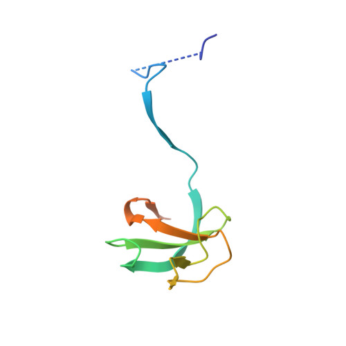
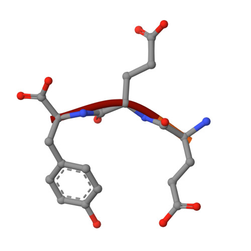
Crystal structure of the A49M mutant CAP-Gly domain of human Dynactin-1 (p150-Glued) in complex with human EB1 C-terminal hexapeptide


Experimental Data Snapshot

  • Method: X-RAY DIFFRACTION
  • Resolution: 2.03 Å
  • R-Value Free: 0.223 
  • R-Value Work: 0.183 
  • R-Value Observed: 0.185 

wwPDB Validation   3D Report Full Report


This is version 1.5 of the entry. See complete history


Literature

Key interaction modes of dynamic +TIP networks.

Honnappa, S.Okhrimenko, O.Jaussi, R.Jawhari, H.Jelesarov, I.Winkler, F.K.Steinmetz, M.O.

(2006) Mol Cell 23: 663-671

  • DOI: https://doi.org/10.1016/j.molcel.2006.07.013
  • Primary Citation of Related Structures:  
    2HKN, 2HKQ, 2HL3, 2HL5

  • PubMed Abstract: 

    Dynamic microtubule plus-end tracking protein (+TIP) networks are implicated in all functions of microtubules, but the molecular determinants of their interactions are largely unknown. Here, we have explored key binding modes of +TIPs by analyzing the interactions between selected CAP-Gly, EB-like, and carboxy-terminal EEY/F-COO(-) sequence motifs. X-ray crystallography and biophysical binding studies demonstrate that the beta2-beta3 loop of CAP-Gly domains determines EB-like motif binding specificity. They further show how CAP-Gly domains serve as recognition domains for EEY/F-COO(-) motifs, which represent characteristic and functionally important sequence elements in EB, CLIP-170, and alpha-tubulin. Our findings provide a molecular basis for understanding the modular interaction modes between alpha-tubulin, CLIPs, EB proteins, and the dynactin-dynein motor complex and suggest that multiple low-affinity binding sites in different combinations control dynamic +TIP networks at microtubule ends. They further offer insights into the structural consequences of genetic CAP-Gly domain defects found in severe human disorders.


  • Organizational Affiliation

    Biomolecular Research, Structural Biology, Paul Scherrer Institut, CH-5232 Villigen PSI, Switzerland.


Macromolecules
Find similar proteins by:  (by identity cutoff)  |  3D Structure
Entity ID: 1
MoleculeChains Sequence LengthOrganismDetailsImage
Dynactin-1
A, B
97Homo sapiensMutation(s): 1 
Gene Names: DCTN1
UniProt & NIH Common Fund Data Resources
Find proteins for Q14203 (Homo sapiens)
Explore Q14203 
Go to UniProtKB:  Q14203
PHAROS:  Q14203
GTEx:  ENSG00000204843 
Entity Groups  
Sequence Clusters30% Identity50% Identity70% Identity90% Identity95% Identity100% Identity
UniProt GroupQ14203
Sequence Annotations
Expand
  • Reference Sequence

Find similar proteins by:  Sequence   |   3D Structure  

Entity ID: 2
MoleculeChains Sequence LengthOrganismDetailsImage
Microtubule-associated protein RP/EB family member 16N/AMutation(s): 0 
UniProt & NIH Common Fund Data Resources
Find proteins for Q15691 (Homo sapiens)
Explore Q15691 
Go to UniProtKB:  Q15691
PHAROS:  Q15691
GTEx:  ENSG00000101367 
Entity Groups  
UniProt GroupQ15691
Sequence Annotations
Expand
  • Reference Sequence
Experimental Data & Validation

Experimental Data

  • Method: X-RAY DIFFRACTION
  • Resolution: 2.03 Å
  • R-Value Free: 0.223 
  • R-Value Work: 0.183 
  • R-Value Observed: 0.185 
  • Space Group: P 21 21 21
Unit Cell:
Length ( Å )Angle ( ˚ )
a = 43.062α = 90
b = 55.068β = 90
c = 66.221γ = 90
Software Package:
Software NamePurpose
REFMACrefinement
XDSdata reduction
XSCALEdata scaling
MOLREPphasing

Structure Validation

View Full Validation Report


Entry History 

Deposition Data

Revision History  (Full details and data files)

  • Version 1.0: 2006-09-12
    Type: Initial release
  • Version 1.1: 2008-05-01
    Changes: Version format compliance
  • Version 1.2: 2011-07-13
    Changes: Version format compliance
  • Version 1.3: 2015-07-29
    Changes: Database references, Structure summary
  • Version 1.4: 2021-10-20
    Changes: Database references
  • Version 1.5: 2024-02-14
    Changes: Data collection