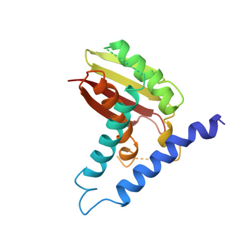
2BJN

X-ray Structure of human TPC6


Experimental Data Snapshot

  • Method: X-RAY DIFFRACTION
  • Resolution: 1.70 Å
  • R-Value Free: 0.209 
  • R-Value Work: 0.175 
  • R-Value Observed: 0.177 

wwPDB Validation   3D Report Full Report


This is version 1.1 of the entry. See complete history


Literature

The Structure of the Trapp Subunit Tpc6 Suggests a Model for a Trapp Subcomplex.

Kummel, D.Mueller, J.J.Roske, Y.Misselwitz, R.Bussow, K.Heinemann, U.

(2005) EMBO Rep 6: 787

  • DOI: https://doi.org/10.1038/sj.embor.7400463
  • Primary Citation of Related Structures:  
    2BJN

  • PubMed Abstract: 

    The TRAPP (transport protein particle) complexes are tethering complexes that have an important role at the different steps of vesicle transport. Recently, the crystal structures of the TRAPP subunits SEDL and BET3 have been determined, and we present here the 1.7 Angstroms crystal structure of human TPC6, a third TRAPP subunit. The protein adopts an alpha/beta-plait topology and forms a dimer. In spite of low sequence similarity, the structure of TPC6 strikingly resembles that of BET3. The similarity is especially prominent at the dimerization interfaces of the proteins. This suggests heterodimerization of TPC6 and BET3, which is shown by in vitro and in vivo association studies. Together with TPC5, another TRAPP subunit, TPC6 and BET3 are supposed to constitute a family of paralogous proteins with closely similar three-dimensional structures but little sequence similarity among its members.


  • Organizational Affiliation

    Max-Delbrück Center for Molecular Medicine, Berlin, Germany.


Macromolecules
Find similar proteins by:  (by identity cutoff)  |  3D Structure
Entity ID: 1
MoleculeChains Sequence LengthOrganismDetailsImage
TRAFFICKING PROTEIN PARTICLE COMPLEX SUBUNIT 6B
A, B
160Homo sapiensMutation(s): 0 
UniProt & NIH Common Fund Data Resources
Find proteins for Q86SZ2 (Homo sapiens)
Explore Q86SZ2 
Go to UniProtKB:  Q86SZ2
PHAROS:  Q86SZ2
GTEx:  ENSG00000182400 
Entity Groups  
Sequence Clusters30% Identity50% Identity70% Identity90% Identity95% Identity100% Identity
UniProt GroupQ86SZ2
Sequence Annotations
Expand
  • Reference Sequence
Small Molecules
Ligands 2 Unique
IDChains Name / Formula / InChI Key2D Diagram3D Interactions
SO4
Query on SO4

Download Ideal Coordinates CCD File 
C [auth A],
D [auth A],
G [auth B],
H [auth B],
I [auth B]
SULFATE ION
O4 S
QAOWNCQODCNURD-UHFFFAOYSA-L
GOL
Query on GOL

Download Ideal Coordinates CCD File 
E [auth A],
F [auth A],
J [auth B],
K [auth B]
GLYCEROL
C3 H8 O3
PEDCQBHIVMGVHV-UHFFFAOYSA-N
Experimental Data & Validation

Experimental Data

  • Method: X-RAY DIFFRACTION
  • Resolution: 1.70 Å
  • R-Value Free: 0.209 
  • R-Value Work: 0.175 
  • R-Value Observed: 0.177 
  • Space Group: C 1 2 1
Unit Cell:
Length ( Å )Angle ( ˚ )
a = 99.423α = 90
b = 57.005β = 117.16
c = 60.406γ = 90
Software Package:
Software NamePurpose
REFMACrefinement
HKL-2000data reduction
HKL-2000data scaling
SOLVE/RESOLVEphasing

Structure Validation

View Full Validation Report


Entry History 

Deposition Data

Revision History  (Full details and data files)

  • Version 1.0: 2005-07-20
    Type: Initial release
  • Version 1.1: 2011-07-13
    Changes: Advisory, Version format compliance