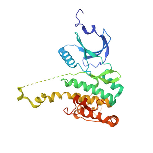
2AC5

Structure of human Mnk2 Kinase Domain mutant D228G


Experimental Data Snapshot

  • Method: X-RAY DIFFRACTION
  • Resolution: 3.20 Å
  • R-Value Free: 0.306 
  • R-Value Work: 0.238 
  • R-Value Observed: 0.306 

wwPDB Validation   3D Report Full Report


This is version 1.4 of the entry. See complete history


Literature

Crystal structures of the Mnk2 kinase domain reveal an inhibitory conformation and a zinc binding site.

Jauch, R.Jakel, S.Netter, C.Schreiter, K.Aicher, B.Jackle, H.Wahl, M.C.

(2005) Structure 13: 1559-1568

  • DOI: https://doi.org/10.1016/j.str.2005.07.013
  • Primary Citation of Related Structures:  
    2AC3, 2AC5

  • PubMed Abstract: 

    Human mitogen-activated protein kinases (MAPK)-interacting kinases 1 and 2 (Mnk1 and Mnk2) target the translational machinery by phosphorylation of the eukaryotic initiation factor 4E (eIF4E). Here, we present the 2.1 A crystal structure of a nonphosphorylated Mnk2 fragment that encompasses the kinase domain. The results show Mnk-specific features such as a zinc binding motif and an atypical open conformation of the activation segment. In addition, the ATP binding pocket contains an Asp-Phe-Asp (DFD) in place of the canonical magnesium binding Asp-Phe-Gly (DFG) motif. The phenylalanine of this motif sticks into the ATP binding pocket and blocks ATP binding as observed with inhibitor bound and, thus, inactive p38 kinase. Replacement of the DFD by the canonical DFG motif affects the conformation of Mnk2, but not ATP binding and kinase activity. The results suggest that the ATP binding pocket and the activation segment of Mnk2 require conformational switches to provide kinase activity.


  • Organizational Affiliation

    Max-Planck-Institut für Biophysikalische Chemie, Abteilung Molekulare Entwicklungsbiologie, Am Fassberg 11, 37077 Göttingen, Germany.


Macromolecules
Find similar proteins by:  (by identity cutoff)  |  3D Structure
Entity ID: 1
MoleculeChains Sequence LengthOrganismDetailsImage
MAP kinase-interacting serine/threonine kinase 2316Homo sapiensMutation(s): 1 
EC: 2.7.1.37 (PDB Primary Data), 2.7.11.1 (UniProt)
UniProt & NIH Common Fund Data Resources
Find proteins for Q9HBH9 (Homo sapiens)
Explore Q9HBH9 
Go to UniProtKB:  Q9HBH9
PHAROS:  Q9HBH9
GTEx:  ENSG00000099875 
Entity Groups  
Sequence Clusters30% Identity50% Identity70% Identity90% Identity95% Identity100% Identity
UniProt GroupQ9HBH9
Sequence Annotations
Expand
  • Reference Sequence
Small Molecules
Ligands 1 Unique
IDChains Name / Formula / InChI Key2D Diagram3D Interactions
ZN
Query on ZN

Download Ideal Coordinates CCD File 
B [auth A]ZINC ION
Zn
PTFCDOFLOPIGGS-UHFFFAOYSA-N
Experimental Data & Validation

Experimental Data

  • Method: X-RAY DIFFRACTION
  • Resolution: 3.20 Å
  • R-Value Free: 0.306 
  • R-Value Work: 0.238 
  • R-Value Observed: 0.306 
  • Space Group: P 32 2 1
Unit Cell:
Length ( Å )Angle ( ˚ )
a = 104.646α = 90
b = 104.646β = 90
c = 73.363γ = 120
Software Package:
Software NamePurpose
DENZOdata reduction
SCALEPACKdata scaling
MOLREPphasing
CNSrefinement

Structure Validation

View Full Validation Report


Entry History 

Deposition Data

Revision History  (Full details and data files)

  • Version 1.0: 2005-10-04
    Type: Initial release
  • Version 1.1: 2008-04-30
    Changes: Version format compliance
  • Version 1.2: 2011-07-13
    Changes: Version format compliance
  • Version 1.3: 2021-11-10
    Changes: Database references, Derived calculations
  • Version 1.4: 2024-05-29
    Changes: Data collection