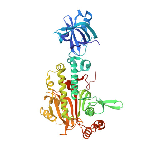
1X54

Crystal structure of asparaginyl-tRNA synthetase from Pyrococcus horikoshii complexed with asparaginyl-adenylate


Experimental Data Snapshot

  • Method: X-RAY DIFFRACTION
  • Resolution: 1.45 Å
  • R-Value Free: 0.192 
  • R-Value Work: 0.168 
  • R-Value Observed: 0.169 

Starting Model: experimental
View more details

wwPDB Validation   3D Report Full Report


Ligand Structure Quality Assessment 


This is version 1.3 of the entry. See complete history


Literature

Structural Basis of the Water-assisted Asparagine Recognition by Asparaginyl-tRNA Synthetase.

Iwasaki, W.Sekine, S.Kuroishi, C.Kuramitsu, S.Shirouzu, M.Yokoyama, S.

(2006) J Mol Biol 360: 329-342

  • DOI: https://doi.org/10.1016/j.jmb.2006.04.068
  • Primary Citation of Related Structures:  
    1X54, 1X55, 1X56

  • PubMed Abstract: 

    Asparaginyl-tRNA synthetase (AsnRS) is a member of the class-II aminoacyl-tRNA synthetases, and is responsible for catalyzing the specific aminoacylation of tRNA(Asn) with asparagine. Here, the crystal structure of AsnRS from Pyrococcus horikoshii, complexed with asparaginyl-adenylate (Asn-AMP), was determined at 1.45 A resolution, and those of free AsnRS and AsnRS complexed with an Asn-AMP analog (Asn-SA) were solved at 1.98 and 1.80 A resolutions, respectively. All of the crystal structures have many solvent molecules, which form a network of hydrogen-bonding interactions that surrounds the entire AsnRS molecule. In the AsnRS/Asn-AMP complex (or the AsnRS/Asn-SA), one side of the bound Asn-AMP (or Asn-SA) is completely covered by the solvent molecules, which complement the binding site. In particular, two of these water molecules were found to interact directly with the asparagine amide and carbonyl groups, respectively, and to contribute to the formation of a pocket highly complementary to the asparagine side-chain. Thus, these two water molecules appear to play a key role in the strict recognition of asparagine and the discrimination against aspartic acid by the AsnRS. This water-assisted asparagine recognition by the AsnRS strikingly contrasts with the fact that the aspartic acid recognition by the closely related aspartyl-tRNA synthetase is achieved exclusively through extensive interactions with protein amino acid residues. Furthermore, based on a docking model of AsnRS and tRNA, a single arginine residue (Arg83) in the AsnRS was postulated to be involved in the recognition of the third position of the tRNA(Asn) anticodon (U36). We performed a mutational analysis of this particular arginine residue, and confirmed its significance in the tRNA recognition.


  • Organizational Affiliation

    Department of Biophysics and Biochemistry, Graduate School of Science, The University of Tokyo, 7-3-1 Hongo, Bunkyo-ku, Tokyo 113-0033, Japan.


Macromolecules
Find similar proteins by:  (by identity cutoff)  |  3D Structure
Entity ID: 1
MoleculeChains Sequence LengthOrganismDetailsImage
Asparaginyl-tRNA synthetase434Pyrococcus horikoshiiMutation(s): 0 
EC: 6.1.1.22
UniProt
Find proteins for O57980 (Pyrococcus horikoshii (strain ATCC 700860 / DSM 12428 / JCM 9974 / NBRC 100139 / OT-3))
Explore O57980 
Go to UniProtKB:  O57980
Entity Groups  
Sequence Clusters30% Identity50% Identity70% Identity90% Identity95% Identity100% Identity
UniProt GroupO57980
Sequence Annotations
Expand
  • Reference Sequence
Small Molecules
Ligands 3 Unique
IDChains Name / Formula / InChI Key2D Diagram3D Interactions
4AD
Query on 4AD

Download Ideal Coordinates CCD File 
C [auth A]4-AMINO-1,4-DIOXOBUTAN-2-AMINIUM ADENOSINE-5'-MONOPHOSPHATE
C14 H21 N7 O9 P
BZHVCXQAFQWXPZ-VWJPMABRSA-O
MPD
Query on MPD

Download Ideal Coordinates CCD File 
D [auth A](4S)-2-METHYL-2,4-PENTANEDIOL
C6 H14 O2
SVTBMSDMJJWYQN-YFKPBYRVSA-N
MG
Query on MG

Download Ideal Coordinates CCD File 
B [auth A]MAGNESIUM ION
Mg
JLVVSXFLKOJNIY-UHFFFAOYSA-N
Experimental Data & Validation

Experimental Data

  • Method: X-RAY DIFFRACTION
  • Resolution: 1.45 Å
  • R-Value Free: 0.192 
  • R-Value Work: 0.168 
  • R-Value Observed: 0.169 
  • Space Group: C 1 2 1
Unit Cell:
Length ( Å )Angle ( ˚ )
a = 126.071α = 90
b = 68.221β = 117.18
c = 75.706γ = 90
Software Package:
Software NamePurpose
REFMACrefinement
HKL-2000data reduction
SCALEPACKdata scaling
MOLREPphasing

Structure Validation

View Full Validation Report


Ligand Structure Quality Assessment 


Entry History 

Deposition Data

Revision History  (Full details and data files)

  • Version 1.0: 2006-05-23
    Type: Initial release
  • Version 1.1: 2008-04-30
    Changes: Version format compliance
  • Version 1.2: 2011-07-13
    Changes: Derived calculations, Version format compliance
  • Version 1.3: 2023-10-25
    Changes: Data collection, Database references, Derived calculations, Refinement description