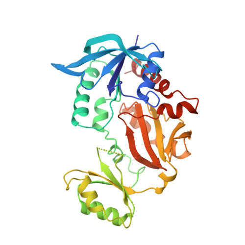
Three-dimensional structure of the glutathione synthetase from Escherichia coli B at 2.0 A resolution.
Yamaguchi, H., Kato, H., Hata, Y., Nishioka, T., Kimura, A., Oda, J., Katsube, Y.(1993) J Mol Biol 229: 1083-1100
- PubMed: 8445637
- DOI: https://doi.org/10.1006/jmbi.1993.1106
- Primary Citation of Related Structures:
1GLV - PubMed Abstract:
Glutathione synthetase (gamma-L-glutamyl-L-cysteine: glycine ligase (ADP-forming) EC 6.3.2.3: GSHase) catalyzes the synthesis of glutathione from gamma-L-glutamyl-L-cysteine and Gly in the presence of ATP. The enzyme from Escherichia coli is a tetramer with four identical subunits of 316 amino acid residues. The crystal structure of the enzyme has been determined by isomorphous replacement and refined to a 2.0 A resolution. Two regions, Gly164 to Gly167 and Ile226 to Arg241, are invisible on the electron density map. The refined model of the subunit includes 296 amino acid residues and 107 solvent molecules. The crystallographic R-factor is 18.6% for 17.914 reflections with F > 3 sigma between 6.0 A and 2.0 A. The structure consists of three domains: the N-terminal, central, and C-terminal domains. In the tetrameric molecule, two subunits that are in close contact form a tight dimer, two tight dimers forming a tetramer with two solvent regions. The ATP molecule is located in the cleft between the central and C-terminal domains. The ATP binding site is surrounded by two sets of the structural motif that belong to those respective domains. Each motif consists of an anti-parallel beta-sheet and a glycine-rich loop.
Organizational Affiliation:
Institute for Protein Research Osaka University, Japan.


