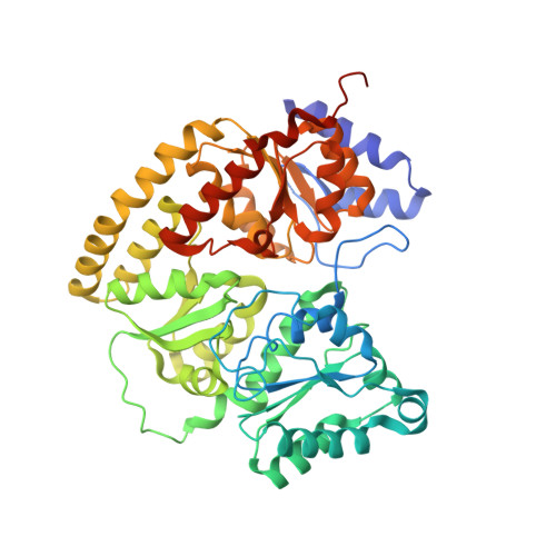
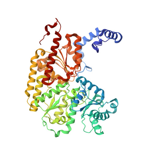
1FP4

CRYSTAL STRUCTURE OF THE ALPHA-H195Q MUTANT OF NITROGENASE


Experimental Data Snapshot

  • Method: X-RAY DIFFRACTION
  • Resolution: 2.50 Å
  • R-Value Free: 0.240 
  • R-Value Work: 0.184 

wwPDB Validation   3D Report Full Report


This is version 1.5 of the entry. See complete history


Literature

Mechanistic features and structure of the nitrogenase alpha-Gln195 MoFe protein

Sorlie, M.Christiansen, J.Lemon, B.J.Peters, J.W.Dean, D.R.Hales, B.J.

(2001) Biochemistry 40: 1540-1549

  • DOI: https://doi.org/10.1021/bi0013997
  • Primary Citation of Related Structures:  
    1FP4

  • PubMed Abstract: 

    EPR signals observed under CO and C(2)H(2) during nitrogenase turnover were investigated for the alpha-Gln(195) MoFe protein, an altered form for which the alpha-His(195) residue has been substituted by glutamine. Under CO, samples show S = 1/2 hi- and lo-CO EPR signals identical to those recognized for the wild-type protein, whereas the S = 3/2 signals generated under high CO/high flux conditions differ. Previous work has revealed that the EPR spectrum generated under C(2)H(2) exhibits a signal (S(EPR1)) originating from the FeMo-cofactor having two or more bound C(2)H(2) adducts and a second signal (S(EPR2)) arising from a radical species [Sørlie, M., Christiansen, J., Dean, D. R., and Hales, B. J. (1999) J. Am. Chem. Soc. 121, 9457-9458]. Pressure-dependent studies show that the intensity of these signals has a sigmoidal dependency at low pressures and maximized at 0.1 atm C(2)H(2) with a subsequent decrease in steady-state intensity at higher pressures. Analogous signals are not recognized for the wild-type MoFe protein. Analysis of the principal g-factors of S(EPR2) suggests that it either represents an unusual metal cluster or is a carboxylate centered radical possibly originating from homocitrate. Both S(EPR1) and S(EPR2) exhibit similar relaxation properties that are atypical for S = 1/2 signals originating from Fe-S clusters or radicals and indicate a coupled relaxation pathway. The alpha-Gln(195) MoFe protein also exhibits these signals when incubated under turnover conditions in the presence of C(2)H(4). Under these conditions, additional inflections in the g 4-6 region assigned to ground-state transitions of an S = 3/2 spin system are also recognized and assigned to turnover states of the MoFe protein without C(2)H(4) bound. The structure of alpha-Gln(195) was crystallographically determined and found to be virtually identical to that of the wild-type MoFe protein except for replacement of an NuH-S hydrogen bond interaction between FeMo-cofactor and the imidazole side chain of alpha-His(195) by an analogous interaction involving Gln.


  • Organizational Affiliation

    Department of Chemistry, Louisiana State University, Baton Rouge, LA 70803-1804, USA.


Macromolecules
Find similar proteins by:  (by identity cutoff)  |  3D Structure
Entity ID: 1
MoleculeChains Sequence LengthOrganismDetailsImage
NITROGENASE MOLYBDENUM-IRON PROTEIN ALPHA CHAIN
A, C
492Azotobacter vinelandiiMutation(s): 1 
EC: 1.18.6.1
UniProt
Find proteins for P07328 (Azotobacter vinelandii)
Explore P07328 
Go to UniProtKB:  P07328
Entity Groups  
Sequence Clusters30% Identity50% Identity70% Identity90% Identity95% Identity100% Identity
UniProt GroupP07328
Sequence Annotations
Expand
  • Reference Sequence
Find similar proteins by:  (by identity cutoff)  |  3D Structure
Entity ID: 2
MoleculeChains Sequence LengthOrganismDetailsImage
NITROGENASE MOLYBDENUM-IRON PROTEIN BETA CHAIN
B, D
523Azotobacter vinelandiiMutation(s): 0 
EC: 1.18.6.1
UniProt
Find proteins for P07329 (Azotobacter vinelandii)
Explore P07329 
Go to UniProtKB:  P07329
Entity Groups  
Sequence Clusters30% Identity50% Identity70% Identity90% Identity95% Identity100% Identity
UniProt GroupP07329
Sequence Annotations
Expand
  • Reference Sequence
Small Molecules
Ligands 4 Unique
IDChains Name / Formula / InChI Key2D Diagram3D Interactions
CFM
Query on CFM

Download Ideal Coordinates CCD File 
F [auth A],
J [auth C]
FE-MO-S CLUSTER
Fe7 Mo S9
UZRXIPMKRKMLQF-UHFFFAOYSA-N
CLP
Query on CLP

Download Ideal Coordinates CCD File 
G [auth A],
K [auth C]
FE-S CLUSTER
Fe8 S8
MJXAKWFPOPRKKV-UHFFFAOYSA-N
HCA
Query on HCA

Download Ideal Coordinates CCD File 
E [auth A],
I [auth C]
3-HYDROXY-3-CARBOXY-ADIPIC ACID
C7 H10 O7
XKJVEVRQMLKSMO-SSDOTTSWSA-N
CA
Query on CA

Download Ideal Coordinates CCD File 
H [auth B],
L [auth D]
CALCIUM ION
Ca
BHPQYMZQTOCNFJ-UHFFFAOYSA-N
Experimental Data & Validation

Experimental Data

  • Method: X-RAY DIFFRACTION
  • Resolution: 2.50 Å
  • R-Value Free: 0.240 
  • R-Value Work: 0.184 
  • Space Group: P 1 21 1
Unit Cell:
Length ( Å )Angle ( ˚ )
a = 107.2α = 90
b = 130.2β = 111.2
c = 80.4γ = 90
Software Package:
Software NamePurpose
SCALEPACKdata scaling
AMoREphasing
X-PLORrefinement

Structure Validation

View Full Validation Report


Entry History 

Deposition Data

Revision History  (Full details and data files)

  • Version 1.0: 2002-11-20
    Type: Initial release
  • Version 1.1: 2008-04-27
    Changes: Version format compliance
  • Version 1.2: 2011-07-13
    Changes: Version format compliance
  • Version 1.3: 2017-10-04
    Changes: Refinement description
  • Version 1.4: 2021-11-10
    Changes: Database references, Derived calculations
  • Version 1.5: 2024-05-29
    Changes: Data collection