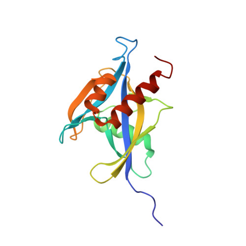
1XSB

Structure of the nudix enzyme AP4A hydrolase from homo sapiens (E63A mutant) in complex with ATP. No ATP restraints included


Experimental Data Snapshot

  • Method: SOLUTION NMR
  • Conformers Calculated: 100 
  • Conformers Submitted: 39 
  • Selection Criteria: target function 

wwPDB Validation   3D Report Full Report


This is version 1.4 of the entry. See complete history


Literature

Structure and Substrate-binding Mechanism of Human Ap4A Hydrolase

Swarbrick, J.D.Buyya, S.Gunawardana, D.Gayler, K.R.McLennan, A.G.Gooley, P.R.

(2005) J Biol Chem 280: 8471-8481

  • DOI: https://doi.org/10.1074/jbc.M412318200
  • Primary Citation of Related Structures:  
    1XSA, 1XSB, 1XSC

  • PubMed Abstract: 

    Asymmetric diadenosine 5',5'''-P(1),P(4)-tetraphosphate (Ap(4)A) hydrolases play a major role in maintaining homeostasis by cleaving the metabolite diadenosine tetraphosphate (Ap(4)A) back into ATP and AMP. The NMR solution structures of the 17-kDa human asymmetric Ap(4)A hydrolase have been solved in both the presence and absence of the product ATP. The adenine moiety of the nucleotide predominantly binds in a ring stacking arrangement equivalent to that observed in the x-ray structure of the homologue from Caenorhabditis elegans. The binding site is, however, markedly divergent to that observed in the plant/pathogenic bacteria class of enzymes, opening avenues for the exploration of specific therapeutics. Binding of ATP induces substantial conformational and dynamic changes that were not observed in the C. elegans structure. In contrast to the C. elegans homologue, important side chains that play a major role in substrate binding do not have to reorient to accommodate the ligand. This may have important implications in the mechanism of substrate recognition in this class of enzymes.


  • Organizational Affiliation

    Department of Biochemistry and Molecular Biology, the University of Melbourne, Parkville, Victoria 3010, Australia.


Macromolecules
Find similar proteins by:  (by identity cutoff)  |  3D Structure
Entity ID: 1
MoleculeChains Sequence LengthOrganismDetailsImage
Bis(5'-nucleosyl)-tetraphosphatase153Homo sapiensMutation(s): 1 
EC: 3.6.1.17
UniProt & NIH Common Fund Data Resources
Find proteins for P50583 (Homo sapiens)
Explore P50583 
Go to UniProtKB:  P50583
PHAROS:  P50583
GTEx:  ENSG00000164978 
Entity Groups  
Sequence Clusters30% Identity50% Identity70% Identity90% Identity95% Identity100% Identity
UniProt GroupP50583
Sequence Annotations
Expand
  • Reference Sequence
Experimental Data & Validation

Experimental Data

  • Method: SOLUTION NMR
  • Conformers Calculated: 100 
  • Conformers Submitted: 39 
  • Selection Criteria: target function 

Structure Validation

View Full Validation Report


Entry History 

Deposition Data

Revision History  (Full details and data files)

  • Version 1.0: 2004-12-21
    Type: Initial release
  • Version 1.1: 2008-04-30
    Changes: Version format compliance
  • Version 1.2: 2011-07-13
    Changes: Version format compliance
  • Version 1.3: 2021-11-10
    Changes: Database references, Derived calculations
  • Version 1.4: 2024-05-29
    Changes: Data collection