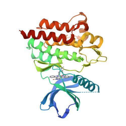
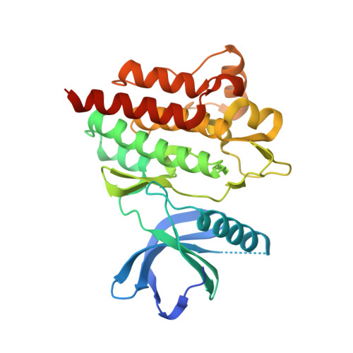
A novel mode of Gleevec binding is revealed by the structure of spleen tyrosine kinase
Atwell, S., Adams, J.M., Badger, J., Buchanan, M.D., Feil, I.K., Froning, K.J., Gao, X., Hendle, J., Keegan, K., Leon, B.C., Muller-Deickmann, H.J., Nienaber, V.L., Noland, B.W., Post, K., Rajashankar, K.R., Ramos, A., Russell, M., Burley, S.K., Buchanan, S.G.(2004) J Biological Chem 279: 55827-55832
- PubMed: 15507431
- DOI: https://doi.org/10.1074/jbc.M409792200
- Primary Citation of Related Structures:
1XBA, 1XBB, 1XBC - PubMed Abstract:
Spleen tyrosine kinase (Syk) is a non-receptor tyrosine kinase required for signaling from immunoreceptors in various hematopoietic cells. Phosphorylation of two tyrosine residues in the activation loop of the Syk kinase catalytic domain is necessary for signaling, a phenomenon typical of tyrosine kinase family members. Syk in vitro enzyme activity, however, does not depend on phosphorylation (activation loop tyrosine --> phenylalanine mutants retain catalytic activity). We have determined the x-ray structure of the unphosphorylated form of the kinase catalytic domain of Syk. The enzyme adopts a conformation of the activation loop typically seen only in activated, phosphorylated tyrosine kinases, explaining why Syk does not require phosphorylation for activation. We also demonstrate that Gleevec (STI-571, Imatinib) inhibits the isolated kinase domains of both unphosphorylated Syk and phosphorylated Abl with comparable potency. Gleevec binds Syk in a novel, compact cis-conformation that differs dramatically from the binding mode observed with unphosphorylated Abl, the more Gleevec-sensitive form of Abl. This finding suggests the existence of two distinct Gleevec binding modes: an extended, trans-conformation characteristic of tight binding to the inactive conformation of a protein kinase and a second compact, cis-conformation characteristic of weaker binding to the active conformation. Finally, the Syk-bound cis-conformation of Gleevec bears a striking resemblance to the rigid structure of the nonspecific, natural product kinase inhibitor staurosporine.
Organizational Affiliation:
Structural GenomiX, Inc., 10505 Roselle Street, San Diego, CA 92121, USA. satwell@stromix.com


