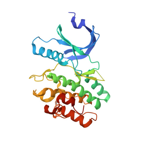
1X8B

Structure of human Wee1A kinase: kinase domain complexed with inhibitor PD0407824


Experimental Data Snapshot

  • Method: X-RAY DIFFRACTION
  • Resolution: 1.81 Å
  • R-Value Free: 0.237 
  • R-Value Work: 0.218 
  • R-Value Observed: 0.219 

wwPDB Validation   3D Report Full Report


Ligand Structure Quality Assessment 


This is version 1.3 of the entry. See complete history


Literature

Structure and inhibition of the human cell cycle checkpoint kinase, Wee1A kinase: an atypical tyrosine kinase with a key role in CDK1 regulation

Squire, C.J.Dickson, J.M.Ivanovic, I.Baker, E.N.

(2005) Structure 13: 541-550

  • DOI: https://doi.org/10.1016/j.str.2004.12.017
  • Primary Citation of Related Structures:  
    1X8B

  • PubMed Abstract: 

    Phosphorylation is critical to regulation of the eukaryotic cell cycle. Entry to mitosis is triggered by the cyclin-dependent kinase CDK1 (Cdc2), which is inactivated during the preceding S and G2 phases by phosphorylation of T14 and Y15. Two homologous kinases, Wee1, which phosphorylates Y15, and Myt1, which phosphorylates both T14 and Y15, mediate this inactivation. We have determined the crystal structure of the catalytic domain of human somatic Wee1 (Wee1A) complexed with an active-site inhibitor at 1.8 A resolution. Although Wee1A is functionally a tyrosine kinase, in sequence and structure it most closely resembles serine/threonine kinases such as Chk1 and cAMP kinases. The crystal structure shows that although the catalytic site closely resembles that of other protein kinases, the activation segment contains Wee1-specific features that maintain it in an active conformation and, together with a key substitution in its glycine-rich loop, help determine its substrate specificity.


  • Organizational Affiliation

    School of Biological Sciences and Centre for Molecular Biodiscovery, University of Auckland, Auckland 1001, New Zealand.


Macromolecules
Find similar proteins by:  (by identity cutoff)  |  3D Structure
Entity ID: 1
MoleculeChains Sequence LengthOrganismDetailsImage
Wee1-like protein kinase289Homo sapiensMutation(s): 0 
EC: 2.7.1.112 (PDB Primary Data), 2.7.10.2 (UniProt)
UniProt & NIH Common Fund Data Resources
Find proteins for P30291 (Homo sapiens)
Explore P30291 
Go to UniProtKB:  P30291
PHAROS:  P30291
GTEx:  ENSG00000166483 
Entity Groups  
Sequence Clusters30% Identity50% Identity70% Identity90% Identity95% Identity100% Identity
UniProt GroupP30291
Sequence Annotations
Expand
  • Reference Sequence
Small Molecules
Ligands 2 Unique
IDChains Name / Formula / InChI Key2D Diagram3D Interactions
824
Query on 824

Download Ideal Coordinates CCD File 
D [auth A]9-HYDROXY-4-PHENYLPYRROLO[3,4-C]CARBAZOLE-1,3(2H,6H)-DIONE
C20 H12 N2 O3
IAUZTOZLTFSMIE-UHFFFAOYSA-N
MG
Query on MG

Download Ideal Coordinates CCD File 
B [auth A],
C [auth A]
MAGNESIUM ION
Mg
JLVVSXFLKOJNIY-UHFFFAOYSA-N
Binding Affinity Annotations 
IDSourceBinding Affinity
824 PDBBind:  1X8B IC50: 97 (nM) from 1 assay(s)
BindingDB:  1X8B IC50: 97 (nM) from 1 assay(s)
Experimental Data & Validation

Experimental Data

  • Method: X-RAY DIFFRACTION
  • Resolution: 1.81 Å
  • R-Value Free: 0.237 
  • R-Value Work: 0.218 
  • R-Value Observed: 0.219 
  • Space Group: P 41 21 2
Unit Cell:
Length ( Å )Angle ( ˚ )
a = 69.782α = 90
b = 69.782β = 90
c = 157.017γ = 90
Software Package:
Software NamePurpose
HKL-2000data collection
SCALEPACKdata scaling
SOLVEphasing
REFMACrefinement
HKL-2000data reduction

Structure Validation

View Full Validation Report


Ligand Structure Quality Assessment 


Entry History 

Deposition Data

Revision History  (Full details and data files)

  • Version 1.0: 2005-06-07
    Type: Initial release
  • Version 1.1: 2008-04-30
    Changes: Version format compliance
  • Version 1.2: 2011-07-13
    Changes: Version format compliance
  • Version 1.3: 2024-03-13
    Changes: Data collection, Database references, Derived calculations