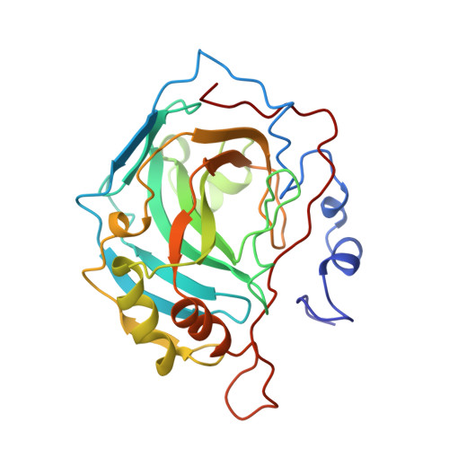
X-ray crystallographic studies of alanine-65 variants of carbonic anhydrase II reveal the structural basis of compromised proton transfer in catalysis.
Scolnick, L.R., Christianson, D.W.(1996) Biochemistry 35: 16429-16434
- PubMed: 8987974
- DOI: https://doi.org/10.1021/bi9617872
- Primary Citation of Related Structures:
1UGA, 1UGB, 1UGC, 1UGD, 1UGE, 1UGF, 1UGG - PubMed Abstract:
The three-dimensional structures of A65F, A65L, A65H, A65T, A65S, and A65G human carbonic anhydrase II (CAII) variants have been solved by X-ray crystallographic methods to probe the importance of residue 65 and the structural implications of its evolutionary drift in the greater family of carbonic anhydrase isozymes. Structure-activity relationships in this series of CAII variants are correlated with those established for other carbonic anhydrase isozymes. We conclude that a bulky side chain at position 65 hinders the formation of an effective solvent bridge between zinc-bound water and H64 and thereby hinders solvent-mediated proton transfer between these two groups [Jackman, J. E., Merz, K. M., Jr., & Fierke, C. A. (1996) Biochemistry 35, 16421-16428]. Despite the introduction of a polar hydroxyl group at this position, smaller side chains such as serine or threonine substituted for A65 do not perturb the formation of a solvent bridge between H64 and zinc-bound solvent. Thus, the evolution of residue 65 size is one factor affecting the trajectory of catalytic proton transfer.
Organizational Affiliation:
Department of Chemistry, University of Pennsylvania, Philadelphia 19104-6323, USA.


