1RYF

Alternative Splicing of Rac1 Generates Rac1b, a Self-activating GTPase


Experimental Data Snapshot

  • Method: X-RAY DIFFRACTION
  • Resolution: 1.75 Å
  • R-Value Free: 0.223 
  • R-Value Work: 0.188 
  • R-Value Observed: 0.190 

Starting Model: experimental
View more details

wwPDB Validation   3D Report Full Report


Ligand Structure Quality Assessment 


This is version 1.3 of the entry. See complete history


Literature

Alternative Splicing of Rac1 Generates Rac1b, a Self-activating GTPase

Fiegen, D.Haeusler, L.C.Blumenstein, L.Herbrand, U.Dvorsky, R.Vetter, I.R.Ahmadian, M.R.

(2004) J Biol Chem 279: 4743-4749

  • DOI: https://doi.org/10.1074/jbc.M310281200
  • Primary Citation of Related Structures:  
    1RYF, 1RYH

  • PubMed Abstract: 

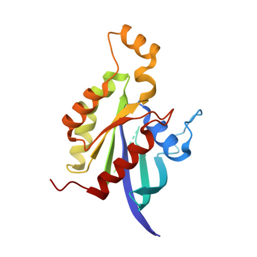
    Rac1b was recently identified in malignant colorectal tumors as an alternative splice variant of Rac1 containing a 19-amino acid insertion next to the switch II region. The structures of Rac1b in the GDP- and the GppNHp-bound forms, determined at a resolution of 1.75 A, reveal that the insertion induces an open switch I conformation and a highly mobile switch II. As a consequence, Rac1b has an accelerated GEF-independent GDP/GTP exchange and an impaired GTP hydrolysis, which is restored partially by GTPase-activating proteins. Interestingly, Rac1b is able to bind the GTPase-binding domain of PAK but not full-length PAK in a GTP-dependent manner, suggesting that the insertion does not completely abolish effector interaction. The presented study provides insights into the structural and biochemical mechanism of a self-activating GTPase.


  • Organizational Affiliation

    Max-Planck-Institut für molekulare Physiologie, Abteilung Strukturelle Biologie, Otto-Hahn-Strasse 11, 44227 Dortmund, Germany.


Macromolecules
Find similar proteins by:  (by identity cutoff)  |  3D Structure
Entity ID: 1
MoleculeChains Sequence LengthOrganismDetailsImage
ras-related C3 botulinum toxin substrate 1 isoform Rac1b
A, B
203Homo sapiensMutation(s): 0 
EC: 3.6.5.2
UniProt & NIH Common Fund Data Resources
Find proteins for P63000 (Homo sapiens)
Explore P63000 
Go to UniProtKB:  P63000
GTEx:  ENSG00000136238 
Entity Groups  
Sequence Clusters30% Identity50% Identity70% Identity90% Identity95% Identity100% Identity
UniProt GroupP63000
Sequence Annotations
Expand
  • Reference Sequence
Small Molecules
Experimental Data & Validation

Experimental Data

  • Method: X-RAY DIFFRACTION
  • Resolution: 1.75 Å
  • R-Value Free: 0.223 
  • R-Value Work: 0.188 
  • R-Value Observed: 0.190 
  • Space Group: P 21 21 21
Unit Cell:
Length ( Å )Angle ( ˚ )
a = 51.64α = 90
b = 78.69β = 90
c = 96.79γ = 90
Software Package:
Software NamePurpose
REFMACrefinement
XDSdata reduction
XDSdata scaling

Structure Validation

View Full Validation Report


Ligand Structure Quality Assessment 


Entry History 

Deposition Data

Revision History  (Full details and data files)

  • Version 1.0: 2004-01-27
    Type: Initial release
  • Version 1.1: 2008-04-29
    Changes: Version format compliance
  • Version 1.2: 2011-07-13
    Changes: Advisory, Refinement description, Version format compliance
  • Version 1.3: 2023-10-25
    Changes: Data collection, Database references, Derived calculations, Refinement description