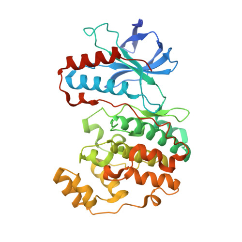
1R39

THE STRUCTURE OF P38ALPHA


Experimental Data Snapshot

  • Method: X-RAY DIFFRACTION
  • Resolution: 2.30 Å
  • R-Value Free: 0.249 
  • R-Value Work: 0.209 
  • R-Value Observed: 0.209 

Starting Model: experimental
View more details

wwPDB Validation   3D Report Full Report


This is version 1.3 of the entry. See complete history


Literature

Lattice stabilization and enhanced diffraction in human p38 alpha crystals by protein engineering.

Patel, S.B.Cameron, P.M.Frantz-Wattley, B.O'Neill, E.Becker, J.W.Scapin, G.

(2004) Biochim Biophys Acta 1696: 67-73

  • DOI: https://doi.org/10.1016/j.bbapap.2003.09.009
  • Primary Citation of Related Structures:  
    1R39, 1R3C

  • PubMed Abstract: 

    Mitogen-activated protein (MAP) kinase p38 alpha is activated in response to environmental stress and cytokines, and plays a significant role in inflammatory responses. For these reasons, it is an important target for the treatment of a wide range of inflammatory and autoimmune diseases. The crystals of p38 alpha that we obtained by published procedures were usually small, quite mosaic, and difficult to reproduce and thus posed a difficulty for the intensive high-resolution studies required for a structure-guided drug discovery approach. Based on crystallographic and biochemical evidences, we prepared a single point mutation of a surface cysteine (C162S) and found that it prevents aggregation and improves the homogeneity and stability of the enzyme. This mutation also facilitates the crystallization process and increases the diffracting power of p38 alpha crystals. Surprisingly, we found that the mutation induces a change in the conformation of a nearby surface loop resulting in stronger lattice interactions, consistent with the improved crystal quality. The mutant protein, because of its improved stability and strengthened lattice interactions, thus provides a significantly improved reagent for use in structure-based drug design for this important disease target.


  • Organizational Affiliation

    Department of Medicinal Chemistry, Merck Research Laboratory, PO Box 2000 RY50-105, Rahway, NJ 07065, USA.


Macromolecules
Find similar proteins by:  (by identity cutoff)  |  3D Structure
Entity ID: 1
MoleculeChains Sequence LengthOrganismDetailsImage
Mitogen-activated protein kinase 14366Homo sapiensMutation(s): 0 
Gene Names: MAPK14CSBP1CSBP2CSBP OR MXI2
EC: 2.7.1.37 (PDB Primary Data), 2.7.11.24 (UniProt)
UniProt & NIH Common Fund Data Resources
Find proteins for Q16539 (Homo sapiens)
Explore Q16539 
Go to UniProtKB:  Q16539
PHAROS:  Q16539
GTEx:  ENSG00000112062 
Entity Groups  
Sequence Clusters30% Identity50% Identity70% Identity90% Identity95% Identity100% Identity
UniProt GroupQ16539
Sequence Annotations
Expand
  • Reference Sequence
Small Molecules
Ligands 1 Unique
IDChains Name / Formula / InChI Key2D Diagram3D Interactions
SO4
Query on SO4

Download Ideal Coordinates CCD File 
B [auth A]SULFATE ION
O4 S
QAOWNCQODCNURD-UHFFFAOYSA-L
Experimental Data & Validation

Experimental Data

  • Method: X-RAY DIFFRACTION
  • Resolution: 2.30 Å
  • R-Value Free: 0.249 
  • R-Value Work: 0.209 
  • R-Value Observed: 0.209 
  • Space Group: P 21 21 21
Unit Cell:
Length ( Å )Angle ( ˚ )
a = 45.181α = 90
b = 84.188β = 90
c = 120.045γ = 90
Software Package:
Software NamePurpose
HKL-2000data collection
SCALEPACKdata scaling
CNSrefinement
HKL-2000data reduction
CNSphasing

Structure Validation

View Full Validation Report


Entry History 

Deposition Data

Revision History  (Full details and data files)

  • Version 1.0: 2004-01-20
    Type: Initial release
  • Version 1.1: 2008-04-29
    Changes: Version format compliance
  • Version 1.2: 2011-07-13
    Changes: Version format compliance
  • Version 1.3: 2023-08-23
    Changes: Data collection, Database references, Derived calculations, Refinement description