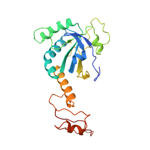
1PRX

HORF6 A NOVEL HUMAN PEROXIDASE ENZYME


Experimental Data Snapshot

  • Method: X-RAY DIFFRACTION
  • Resolution: 2.00 Å
  • R-Value Free: 0.259 
  • R-Value Work: 0.191 
  • R-Value Observed: 0.191 

wwPDB Validation   3D Report Full Report


This is version 1.4 of the entry. See complete history


Literature

Crystal structure of a novel human peroxidase enzyme at 2.0 A resolution.

Choi, H.J.Kang, S.W.Yang, C.H.Rhee, S.G.Ryu, S.E.

(1998) Nat Struct Biol 5: 400-406

  • DOI: https://doi.org/10.1038/nsb0598-400
  • Primary Citation of Related Structures:  
    1PRX

  • PubMed Abstract: 

    Hydrogen peroxide (H2O2) has been implicated recently as an intracellular messenger that affects cellular processes including protein phosphorylation, transcription and apoptosis. A set of novel peroxidases, named peroxiredoxins (Prx), regulate the intracellular concentration of H2O2 by reducing it in the presence of an appropriate electron donor. The crystal structure of a human Prx enzyme, hORF6, reveals that the protein contains two discrete domains and forms a dimer. The N-terminal domain has a thioredoxin fold and the C-terminal domain is used for dimerization. The active site cysteine (Cys 47), which exists as cysteine-sulfenic acid in the crystal, is located at the bottom of a relatively narrow pocket. The positively charged environment surrounding Cys 47 accounts for the peroxidase activity of the enzyme, which contains no redox cofactors.


  • Organizational Affiliation

    Division of Protein Engineering, Korea Research Institute of Bioscience and Biotechnology, KIST, Yusong, Taejon, South Korea.


Macromolecules
Find similar proteins by:  (by identity cutoff)  |  3D Structure
Entity ID: 1
MoleculeChains Sequence LengthOrganismDetailsImage
HORF6
A, B
224Homo sapiensMutation(s): 2 
EC: 3.1.1.4 (UniProt), 2.3.1.23 (UniProt), 1.11.1.27 (UniProt)
UniProt & NIH Common Fund Data Resources
Find proteins for P30041 (Homo sapiens)
Explore P30041 
Go to UniProtKB:  P30041
PHAROS:  P30041
GTEx:  ENSG00000117592 
Entity Groups  
Sequence Clusters30% Identity50% Identity70% Identity90% Identity95% Identity100% Identity
UniProt GroupP30041
Sequence Annotations
Expand
  • Reference Sequence
Small Molecules
Modified Residues  1 Unique
IDChains TypeFormula2D DiagramParent
CSO
Query on CSO
A, B
L-PEPTIDE LINKINGC3 H7 N O3 SCYS
Experimental Data & Validation

Experimental Data

  • Method: X-RAY DIFFRACTION
  • Resolution: 2.00 Å
  • R-Value Free: 0.259 
  • R-Value Work: 0.191 
  • R-Value Observed: 0.191 
  • Space Group: P 1 21 1
Unit Cell:
Length ( Å )Angle ( ˚ )
a = 47.85α = 90
b = 75.17β = 110.21
c = 63.3γ = 90
Software Package:
Software NamePurpose
X-PLORmodel building
X-PLORrefinement
R-AXISdata reduction
R-AXISdata scaling
X-PLORphasing

Structure Validation

View Full Validation Report


Entry History 

Deposition Data

Revision History  (Full details and data files)

  • Version 1.0: 1998-06-17
    Type: Initial release
  • Version 1.1: 2008-03-24
    Changes: Version format compliance
  • Version 1.2: 2011-07-13
    Changes: Version format compliance
  • Version 1.3: 2021-11-03
    Changes: Database references, Derived calculations, Other
  • Version 1.4: 2024-10-30
    Changes: Data collection, Structure summary