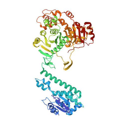
1PL0

Crystal structure of human ATIC in complex with folate-based inhibitor, BW2315U89UC


Experimental Data Snapshot

  • Method: X-RAY DIFFRACTION
  • Resolution: 2.60 Å
  • R-Value Free: 0.275 
  • R-Value Work: 0.213 

Starting Model: experimental
View more details

wwPDB Validation   3D Report Full Report


Ligand Structure Quality Assessment 


This is version 1.3 of the entry. See complete history


Literature

Crystal Structures of Human Bifunctional Enzyme Aminoimidazole-4-carboxamide Ribonucleotide Transformylase/IMP Cyclohydrolase in Complex with Potent Sulfonyl-containing Antifolates.

Cheong, C.G.Wolan, D.W.Greasley, S.E.Horton, P.A.Beardsley, G.P.Wilson, I.A.

(2004) J Biol Chem 279: 18034-18045

  • DOI: https://doi.org/10.1074/jbc.M313691200
  • Primary Citation of Related Structures:  
    1P4R, 1PL0

  • PubMed Abstract: 

    Aminoimidazole-4-carboxamide ribonucleotide (AICAR) transformylase/IMP cyclohydrolase (ATIC) is a bifunctional enzyme with folate-dependent AICAR transformylase and IMP cyclohydrolase activities that catalyzes the last two steps of purine biosynthesis. The AICAR transformylase inhibitors BW1540 and BW2315 are sulfamido-bridged 5,8-dideazafolate analogs with remarkably potent K(i) values of 8 and 6 nm, respectively, compared with most other antifolates. Crystal structures of ATIC at 2.55 and 2.60 A with each inhibitor, in the presence of substrate AICAR, revealed that the sulfonyl groups dominate inhibitor binding and orientation through interaction with the proposed oxyanion hole. These agents then appear to mimic the anionic transition state and now implicate Asn(431') in the reaction mechanism along with previously identified key catalytic residues Lys(266) and His(267). Potent and selective inhibition of the AICAR transformylase active site, compared with other folate-dependent enzymes, should therefore be pursued by further design of sulfonyl-containing antifolates.


  • Organizational Affiliation

    Department of Molecular Biology and The Skaggs Institute for Chemical Biology, The Scripps Research Institute, La Jolla, California 92037, USA.


Macromolecules
Find similar proteins by:  (by identity cutoff)  |  3D Structure
Entity ID: 1
MoleculeChains Sequence LengthOrganismDetailsImage
Bifunctional purine biosynthesis protein PURH
A, B, C, D
592Homo sapiensMutation(s): 0 
Gene Names: ATIC
EC: 2.1.2.3 (PDB Primary Data), 3.5.4.10 (PDB Primary Data)
UniProt & NIH Common Fund Data Resources
Find proteins for P31939 (Homo sapiens)
Explore P31939 
Go to UniProtKB:  P31939
PHAROS:  P31939
GTEx:  ENSG00000138363 
Entity Groups  
Sequence Clusters30% Identity50% Identity70% Identity90% Identity95% Identity100% Identity
UniProt GroupP31939
Sequence Annotations
Expand
  • Reference Sequence
Small Molecules
Ligands 4 Unique
IDChains Name / Formula / InChI Key2D Diagram3D Interactions
BW2
Query on BW2

Download Ideal Coordinates CCD File 
I [auth B],
O [auth D]
N-(4-{[(2-AMINO-4-OXO-3,4-DIHYDROQUINAZOLIN-6-YL)AMINO]SULFONYL}BENZOYL)GLUTAMIC ACID
C20 H19 N5 O8 S
ILTMHHAQXFNQFZ-UHFFFAOYSA-N
XMP
Query on XMP

Download Ideal Coordinates CCD File 
F [auth A],
K [auth C]
XANTHOSINE-5'-MONOPHOSPHATE
C10 H14 N4 O9 P
DCTLYFZHFGENCW-UUOKFMHZSA-O
AMZ
Query on AMZ

Download Ideal Coordinates CCD File 
H [auth B],
M [auth D],
N [auth D]
AMINOIMIDAZOLE 4-CARBOXAMIDE RIBONUCLEOTIDE
C9 H15 N4 O8 P
NOTGFIUVDGNKRI-UUOKFMHZSA-N
K
Query on K

Download Ideal Coordinates CCD File 
E [auth A],
G [auth B],
J [auth C],
L [auth D]
POTASSIUM ION
K
NPYPAHLBTDXSSS-UHFFFAOYSA-N
Binding Affinity Annotations 
IDSourceBinding Affinity
BW2 BindingDB:  1PL0 Ki: 6 (nM) from 1 assay(s)
IC50: 1000 (nM) from 1 assay(s)
PDBBind:  1PL0 Ki: 6 (nM) from 1 assay(s)
Experimental Data & Validation

Experimental Data

  • Method: X-RAY DIFFRACTION
  • Resolution: 2.60 Å
  • R-Value Free: 0.275 
  • R-Value Work: 0.213 
  • Space Group: P 1 21 1
Unit Cell:
Length ( Å )Angle ( ˚ )
a = 77.12α = 90
b = 92.97β = 91.19
c = 178.49γ = 90
Software Package:
Software NamePurpose
SCALEPACKdata scaling
AMoREphasing
CNSrefinement

Structure Validation

View Full Validation Report


Ligand Structure Quality Assessment 


Entry History 

Deposition Data

Revision History  (Full details and data files)

  • Version 1.0: 2004-04-27
    Type: Initial release
  • Version 1.1: 2008-04-29
    Changes: Version format compliance
  • Version 1.2: 2011-07-13
    Changes: Version format compliance
  • Version 1.3: 2023-08-16
    Changes: Data collection, Database references, Derived calculations, Refinement description