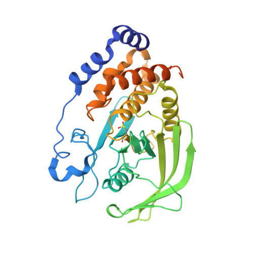
1OEU

Oxidation state of protein tyrosine phosphatase 1B


Experimental Data Snapshot

  • Method: X-RAY DIFFRACTION
  • Resolution: 2.50 Å
  • R-Value Free: 0.270 
  • R-Value Work: 0.196 
  • R-Value Observed: 0.200 

Starting Model: experimental
View more details

wwPDB Validation   3D Report Full Report


This is version 1.4 of the entry. See complete history


Literature

Oxidation state of the active-site cysteine in protein tyrosine phosphatase 1B.

van Montfort, R.L.Congreve, M.Tisi, D.Carr, R.Jhoti, H.

(2003) Nature 423: 773-777

  • DOI: https://doi.org/10.1038/nature01681
  • Primary Citation of Related Structures:  
    1OES, 1OET, 1OEU, 1OEV

  • PubMed Abstract: 

    Protein tyrosine phosphatases regulate signal transduction pathways involving tyrosine phosphorylation and have been implicated in the development of cancer, diabetes, rheumatoid arthritis and hypertension. Increasing evidence suggests that the cellular redox state is involved in regulating tyrosine phosphatase activity through the reversible oxidization of the catalytic cysteine to sulphenic acid (Cys-SOH). But how further oxidation to the irreversible sulphinic (Cys-SO2H) and sulphonic (Cys-SO3H) forms is prevented remains unclear. Here we report the crystal structures of the regulatory sulphenic and irreversible sulphinic and sulphonic acids of protein tyrosine phosphatase 1B (PTP1B), an important enzyme in the negative regulation of the insulin receptor and a therapeutic target in type II diabetes and obesity. We also identify a sulphenyl-amide species that is formed through oxidation of its catalytic cysteine. Formation of the sulphenyl-amide causes large changes in the PTP1B active site, which are reversible by reduction with the cellular reducing agent glutathione. The sulphenyl-amide is a protective intermediate in the oxidative inhibition of PTP1B. In addition, it may facilitate reactivation of PTP1B by biological thiols and signal a unique state of the protein.


  • Organizational Affiliation

    Astex Technology Ltd, 436 Cambridge Science Park, Milton Road, Cambridge CB4 0QA, UK.


Macromolecules
Find similar proteins by:  (by identity cutoff)  |  3D Structure
Entity ID: 1
MoleculeChains Sequence LengthOrganismDetailsImage
PROTEIN-TYROSINE PHOSPHATASE, NON-RECEPTOR TYPE 1321Homo sapiensMutation(s): 0 
EC: 3.1.3.48
UniProt & NIH Common Fund Data Resources
Find proteins for P18031 (Homo sapiens)
Explore P18031 
Go to UniProtKB:  P18031
PHAROS:  P18031
GTEx:  ENSG00000196396 
Entity Groups  
Sequence Clusters30% Identity50% Identity70% Identity90% Identity95% Identity100% Identity
UniProt GroupP18031
Sequence Annotations
Expand
  • Reference Sequence
Small Molecules
Ligands 1 Unique
IDChains Name / Formula / InChI Key2D Diagram3D Interactions
MG
Query on MG

Download Ideal Coordinates CCD File 
B [auth A]MAGNESIUM ION
Mg
JLVVSXFLKOJNIY-UHFFFAOYSA-N
Modified Residues  1 Unique
IDChains TypeFormula2D DiagramParent
CSD
Query on CSD
A
L-PEPTIDE LINKINGC3 H7 N O4 SCYS
Experimental Data & Validation

Experimental Data

  • Method: X-RAY DIFFRACTION
  • Resolution: 2.50 Å
  • R-Value Free: 0.270 
  • R-Value Work: 0.196 
  • R-Value Observed: 0.200 
  • Space Group: P 31 2 1
Unit Cell:
Length ( Å )Angle ( ˚ )
a = 88.914α = 90
b = 88.914β = 90
c = 104.645γ = 120
Software Package:
Software NamePurpose
REFMACrefinement
d*TREKdata reduction
d*TREKdata scaling
AMoREphasing

Structure Validation

View Full Validation Report


Entry History 

Deposition Data

Revision History  (Full details and data files)

  • Version 1.0: 2003-06-12
    Type: Initial release
  • Version 1.1: 2014-11-05
    Changes: Atomic model, Derived calculations, Non-polymer description, Other, Structure summary, Version format compliance
  • Version 1.2: 2017-07-05
    Changes: Data collection
  • Version 1.3: 2018-03-28
    Changes: Database references, Source and taxonomy, Structure summary
  • Version 1.4: 2023-12-13
    Changes: Data collection, Database references, Derived calculations, Refinement description