1MJV

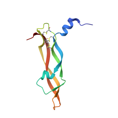
DISULFIDE DEFICIENT MUTANT OF VASCULAR ENDOTHELIAL GROWTH FACTOR A (C51A and C60A)


Experimental Data Snapshot

  • Method: X-RAY DIFFRACTION
  • Resolution: 2.10 Å
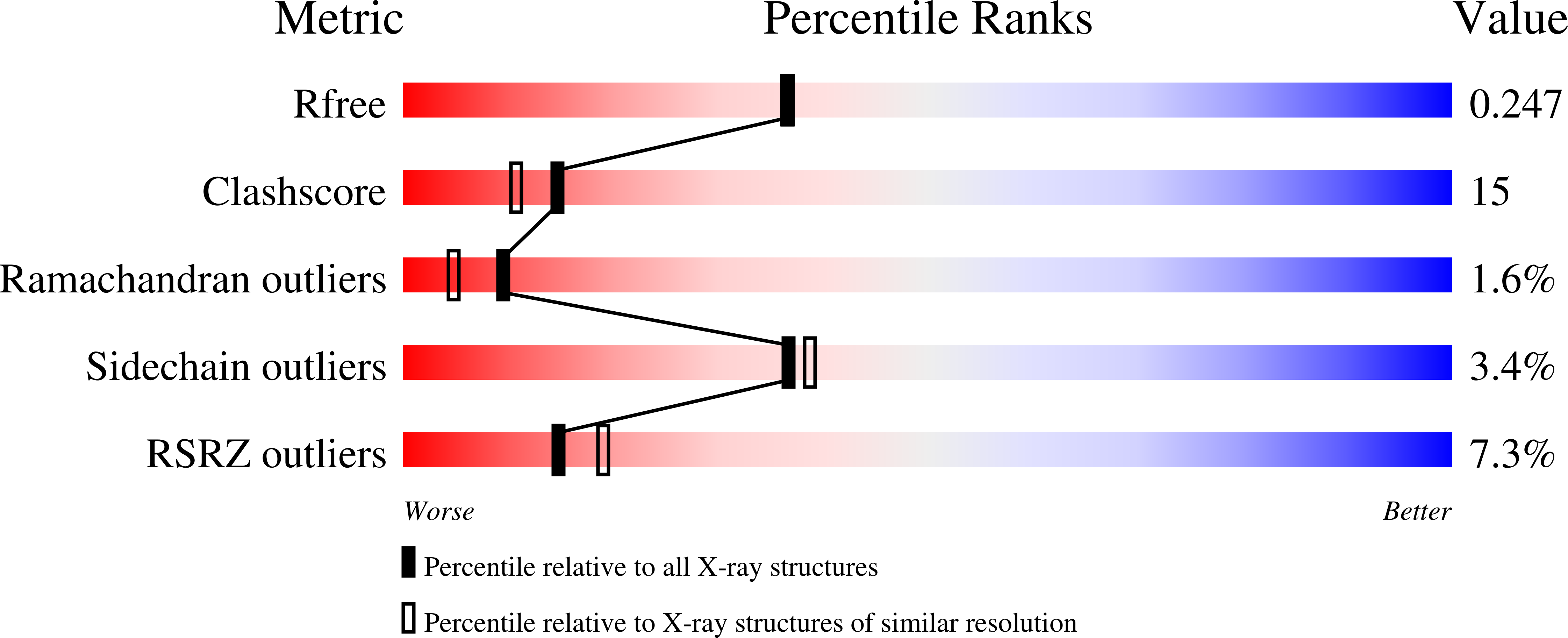
  • R-Value Free: 0.248 
  • R-Value Work: 0.196 
  • R-Value Observed: 0.203 

Starting Model: experimental
View more details

wwPDB Validation   3D Report Full Report


This is version 1.6 of the entry. See complete history


Literature

The cystine knot promotes folding and not thermodynamic stability in vascular endothelial growth factor

Muller, Y.A.Heiring, C.Misselwitz, R.Welfle, K.Welfle, H.

(2002) J Biol Chem 277: 43410-43416

  • DOI: https://doi.org/10.1074/jbc.M206438200
  • Primary Citation of Related Structures:  
    1MJV, 1MKG, 1MKK

  • PubMed Abstract: 

    Cystine knots consist of three intertwined disulfide bridges and are considered major determinants of protein stability in proteins in which they occur. We questioned this function and observed that removal of individual disulfide bridges in human vascular endothelial growth factor (VEGF) does not reduce its thermodynamic stability but reduces its unexpected high thermal stability of 108 degrees C by up to 40 degrees C. In wild-type VEGF (deltaG(u,25)(0) = 5.1 kcal.mol(-1)), the knot is responsible for a large entropic stabilization of TdeltaS(u,25)(0) = -39.3 kcal mol(-1), which is compensated for by a deltaH(u,25)(0) of -34.2 kcal mol(-1). In the disulfide-deficient mutants, this entropic stabilization disappears, but instead of a decrease, we observe an increase in the thermodynamic stability by about 2 kcal.mol(-1). A detailed crystallographic analysis of the mutant structures suggests a role of the cystine knot motif in protein folding rather than in the stabilization of the folded state. When assuming that the sequential order of the disulfide bridge formation is conserved between VEGF and glycoprotein alpha-subunit, the crystal structure of the mutant C61A-C104A, which deviates by a root mean square deviation of more than 2.2 A from wild-type VEGF, identifies a true folding intermediate of VEGF.


  • Organizational Affiliation

    Forschungsgruppe Kristallographie, Max-Delbrück-Centrum für Molekulare Medizin, Berlin, D-13092 Berlin, Germany. y.muller@sussex.ac.uk


Macromolecules
Find similar proteins by:  (by identity cutoff)  |  3D Structure
Entity ID: 1
MoleculeChains Sequence LengthOrganismDetailsImage
Vascular Endothelial Growth Factor A
A, B
96Homo sapiensMutation(s): 2 
UniProt & NIH Common Fund Data Resources
Find proteins for P15692 (Homo sapiens)
Explore P15692 
Go to UniProtKB:  P15692
PHAROS:  P15692
GTEx:  ENSG00000112715 
Entity Groups  
Sequence Clusters30% Identity50% Identity70% Identity90% Identity95% Identity100% Identity
UniProt GroupP15692
Sequence Annotations
Expand
  • Reference Sequence
Experimental Data & Validation

Experimental Data

  • Method: X-RAY DIFFRACTION
  • Resolution: 2.10 Å
  • R-Value Free: 0.248 
  • R-Value Work: 0.196 
  • R-Value Observed: 0.203 
  • Space Group: P 1 21 1
Unit Cell:
Length ( Å )Angle ( ˚ )
a = 29.28α = 90
b = 77.2β = 98.92
c = 55.11γ = 90
Software Package:
Software NamePurpose
MAR345data collection
XDSdata reduction
AMoREphasing
CNSrefinement
XDSdata scaling

Structure Validation

View Full Validation Report


Entry History 

Deposition Data

Revision History  (Full details and data files)

  • Version 1.0: 2002-12-11
    Type: Initial release
  • Version 1.1: 2008-04-28
    Changes: Version format compliance
  • Version 1.2: 2011-07-13
    Changes: Version format compliance
  • Version 1.3: 2017-10-11
    Changes: Refinement description
  • Version 1.4: 2018-04-04
    Changes: Data collection
  • Version 1.5: 2021-10-27
    Changes: Database references
  • Version 1.6: 2024-10-30
    Changes: Data collection, Refinement description, Structure summary