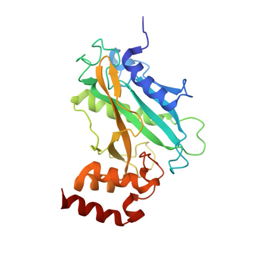
Suppressor of fused regulates Gli activity through a dual binding mechanism
Merchant, M., Vajdos, F.F., Ultsch, M., Maun, H.R., Wendt, U., Cannon, J., Desmarais, W., Lazarus, R.A., de Vos, A.M., de Sauvage, F.J.(2004) Mol Cell Biol 24: 8627-8641
- PubMed: 15367681
- DOI: https://doi.org/10.1128/MCB.24.19.8627-8641.2004
- Primary Citation of Related Structures:
1M1L - PubMed Abstract:
The Hedgehog pathway drives proliferation and differentiation by activating the Gli/Ci family of zinc finger transcription factors. Gli/Ci proteins form Hedgehog signaling complexes with other signaling components, including the kinesin-like protein Costal-2, the serine-threonine kinase Fused, and Suppressor of Fused [Su(fu)]. In these complexes Gli/Ci proteins are regulated by cytoplasmic sequestration, phosphorylation, and proteolysis. Here we characterize structural and functional determinants of Su(fu) required for Gli regulation and show that Su(fu) contains at least two distinct domains: a highly conserved carboxy-terminal region required for binding to the amino-terminal ends of the Gli proteins and a unique amino-terminal domain that binds the carboxy-terminal tail of Gli1. While each domain is capable of binding to different Gli1 regions independently, interactions between Su(fu) and Gli1 at both sites are required for cytoplasmic tethering and repression of Gli1. Furthermore, we have solved the crystal structure of the amino-terminal domain of human Su(fu)(27-268) at 2.65 A resolution. This domain forms a concave pocket with a prominent acidic patch. Mutation at Asp(159) in the acidic patch disrupts Gli1 tethering and repression while not strongly disrupting binding, indicating that the amino-terminal domain of Su(fu) likely impacts Gli binding through a mechanism distinct from that for tethering and repression. These studies provide a structural basis for understanding the function of Su(fu).
Organizational Affiliation:
Department of Molecular Biology, Genentech, Inc., 1 DNA Way, South San Francisco, CA 94080, USA. mmerch@gene.com


