1KN0

Crystal Structure of the human Rad52 protein


Experimental Data Snapshot

  • Method: X-RAY DIFFRACTION
  • Resolution: 2.85 Å
  • R-Value Free: 0.297 
  • R-Value Work: 0.231 
  • R-Value Observed: 0.236 

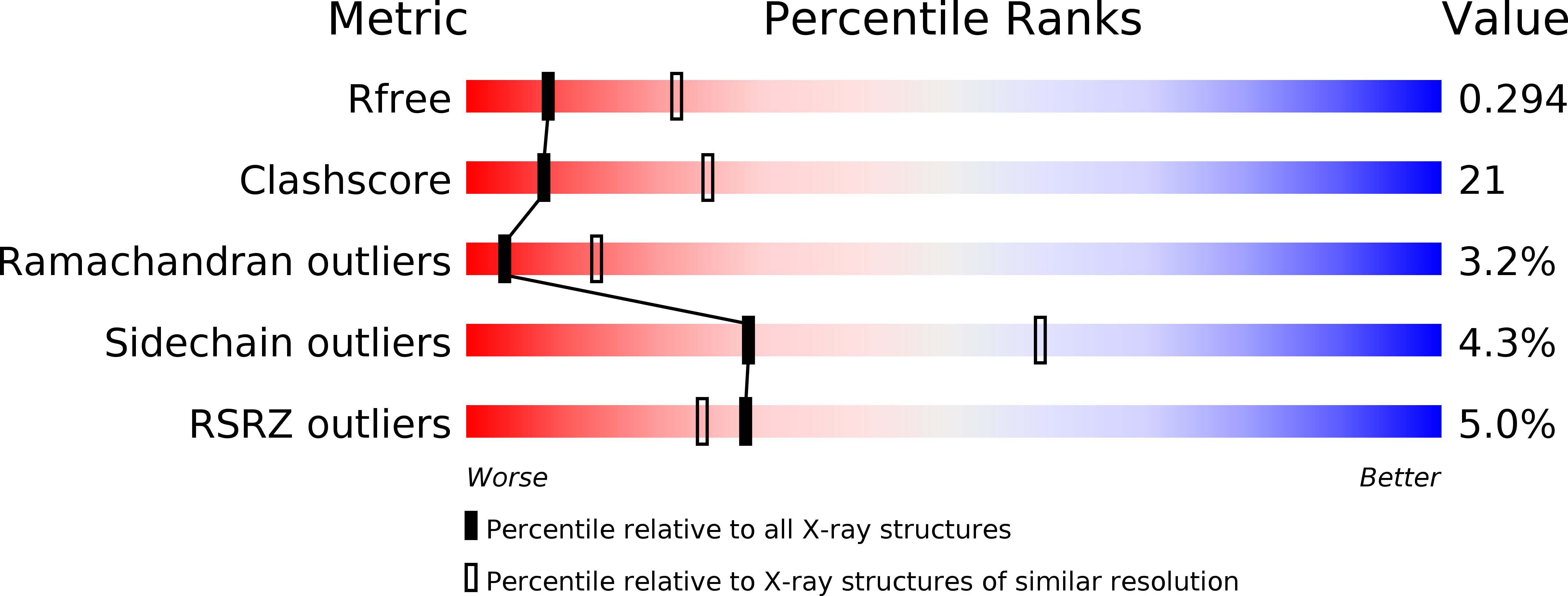
wwPDB Validation   3D Report Full Report


This is version 1.3 of the entry. See complete history


Literature

Crystal structure of the homologous-pairing domain from the human Rad52 recombinase in the undecameric form.

Kagawa, W.Kurumizaka, H.Ishitani, R.Fukai, S.Nureki, O.Shibata, T.Yokoyama, S.

(2002) Mol Cell 10: 359-371

  • DOI: https://doi.org/10.1016/s1097-2765(02)00587-7
  • Primary Citation of Related Structures:  
    1KN0

  • PubMed Abstract: 

    The human Rad52 protein forms a heptameric ring that catalyzes homologous pairing. The N-terminal half of Rad52 is the catalytic domain for homologous pairing, and the ring formed by the domain fragment was reported to be approximately decameric. Splicing variants of Rad52 and a yeast homolog (Rad59) are composed mostly of this domain. In this study, we determined the crystal structure of the homologous-pairing domain of human Rad52 and revealed that the domain forms an undecameric ring. Each monomer has a beta-beta-beta-alpha fold, which consists of highly conserved amino acid residues among Rad52 homologs. A mutational analysis revealed that the amino acid residues located between the beta-beta-beta-alpha fold and the characteristic hairpin loop are essential for ssDNA and dsDNA binding.


  • Organizational Affiliation

    RIKEN Genomic Sciences Center, 1-7-22 Suehiro-cho, Tsurumi, Yokohama, Japan.


Macromolecules
Find similar proteins by:  (by identity cutoff)  |  3D Structure
Entity ID: 1
MoleculeChains Sequence LengthOrganismDetailsImage
Rad52
A, B, C, D, E
A, B, C, D, E, F, G, H, I, J, K
212Homo sapiensMutation(s): 0 
Gene Names: Rad52
UniProt & NIH Common Fund Data Resources
Find proteins for P43351 (Homo sapiens)
Explore P43351 
Go to UniProtKB:  P43351
PHAROS:  P43351
GTEx:  ENSG00000002016 
Entity Groups  
Sequence Clusters30% Identity50% Identity70% Identity90% Identity95% Identity100% Identity
UniProt GroupP43351
Sequence Annotations
Expand
  • Reference Sequence
Experimental Data & Validation

Experimental Data

  • Method: X-RAY DIFFRACTION
  • Resolution: 2.85 Å
  • R-Value Free: 0.297 
  • R-Value Work: 0.231 
  • R-Value Observed: 0.236 
  • Space Group: P 43 21 2
Unit Cell:
Length ( Å )Angle ( ˚ )
a = 145.61α = 90
b = 145.61β = 90
c = 247.53γ = 90
Software Package:
Software NamePurpose
SHARPphasing
CNSrefinement
DENZOdata reduction
SCALEPACKdata scaling

Structure Validation

View Full Validation Report


Entry History 

Revision History  (Full details and data files)

  • Version 1.0: 2002-09-04
    Type: Initial release
  • Version 1.1: 2008-04-27
    Changes: Version format compliance
  • Version 1.2: 2011-07-13
    Changes: Version format compliance
  • Version 1.3: 2024-03-13
    Changes: Data collection, Database references