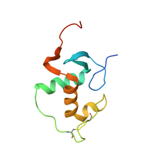
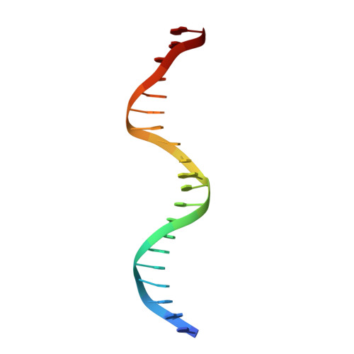
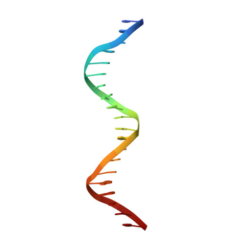
1HLZ

CRYSTAL STRUCTURE OF THE ORPHAN NUCLEAR RECEPTOR REV-ERB(ALPHA) DNA-BINDING DOMAIN BOUND TO ITS COGNATE RESPONSE ELEMENT


Experimental Data Snapshot

  • Method: X-RAY DIFFRACTION
  • Resolution: 2.80 Å
  • R-Value Free: 0.310 
  • R-Value Work: 0.260 
  • R-Value Observed: 0.260 

Starting Model: experimental
View more details

wwPDB Validation   3D Report Full Report


This is version 1.3 of the entry. See complete history


Literature

DNA Deformability as a Recognition Feature in the RevErb Response Element

Sierk, M.L.Zhao, Q.Rastinejad, F.

(2001) Biochemistry 40: 12833-12843

  • DOI: https://doi.org/10.1021/bi011086r
  • Primary Citation of Related Structures:  
    1GA5, 1HLZ

  • PubMed Abstract: 

    Most nuclear receptors recognize the same consensus hexameric sequence, AGGTCA. An important question has been how the various members of this transcription factor family distinguish identity features in these closely related DNA sites. We determined structures from several crystal forms of the RevErb-DNA complex and analyzed the patterns of protein-DNA interactions and DNA distortions. We found a significant and consistent DNA distortion at a TA step directly preceding the first consensus 5'-AGGTCA-3' recognition sequence. Importantly, while this base-pair sequence is associated with RevErb's high-affinity sites, there are no sequence-specific contacts formed with the protein. Our study shows that RevErb relies instead on the intrinsic geometry and flexibility of this TA site to make the required fit between the proteins' independent major groove and minor groove binding interactions, which occur on both sides of the TA step. Our findings extend the description of response element discrimination to include a role for sequence-dependent DNA deformations and suggest how other monomeric members of this superfamily, such as NGFI-B, SF-1, and ROR, could also recognize unique geometric features in their DNA targets.


  • Organizational Affiliation

    Department of Pharmacology, University of Virginia, Charlottesville, Virginia 22908-0735, USA.


Macromolecules

Find similar proteins by:  (by identity cutoff)  |  3D Structure
Entity ID: 3
MoleculeChains Sequence LengthOrganismDetailsImage
ORPHAN NUCLEAR RECEPTOR NR1D1C [auth A],
D [auth B]
94Homo sapiensMutation(s): 0 
UniProt & NIH Common Fund Data Resources
Find proteins for P20393 (Homo sapiens)
Explore P20393 
Go to UniProtKB:  P20393
PHAROS:  P20393
GTEx:  ENSG00000126368 
Entity Groups  
Sequence Clusters30% Identity50% Identity70% Identity90% Identity95% Identity100% Identity
UniProt GroupP20393
Sequence Annotations
Expand
  • Reference Sequence

Find similar nucleic acids by:  Sequence   |   3D Structure  

Entity ID: 1
MoleculeChains LengthOrganismImage
5'-D(*CP*AP*AP*CP*TP*AP*GP*GP*TP*CP*AP*CP*TP*AP*GP*GP*TP*CP*AP*G)-3'A [auth C]20N/A
Sequence Annotations
Expand
  • Reference Sequence

Find similar nucleic acids by:  Sequence   |   3D Structure  

Entity ID: 2
MoleculeChains LengthOrganismImage
5'-D(*CP*TP*GP*AP*CP*CP*TP*AP*GP*TP*GP*AP*CP*CP*TP*AP*GP*TP*TP*G)-3'B [auth D]20N/A
Sequence Annotations
Expand
  • Reference Sequence
Experimental Data & Validation

Experimental Data

  • Method: X-RAY DIFFRACTION
  • Resolution: 2.80 Å
  • R-Value Free: 0.310 
  • R-Value Work: 0.260 
  • R-Value Observed: 0.260 
  • Space Group: P 1
Unit Cell:
Length ( Å )Angle ( ˚ )
a = 35.867α = 84.13
b = 39.603β = 72.09
c = 58.565γ = 70.2
Software Package:
Software NamePurpose
DENZOdata reduction
SCALEPACKdata scaling
AMoREphasing
CNSrefinement

Structure Validation

View Full Validation Report


Entry History 

Deposition Data

Revision History  (Full details and data files)

  • Version 1.0: 2001-11-16
    Type: Initial release
  • Version 1.1: 2008-04-27
    Changes: Version format compliance
  • Version 1.2: 2011-07-13
    Changes: Version format compliance
  • Version 1.3: 2023-08-09
    Changes: Data collection, Database references, Derived calculations, Refinement description