1HCF

Crystal structure of TrkB-d5 bound to neurotrophin-4/5


Experimental Data Snapshot

  • Method: X-RAY DIFFRACTION
  • Resolution: 2.70 Å
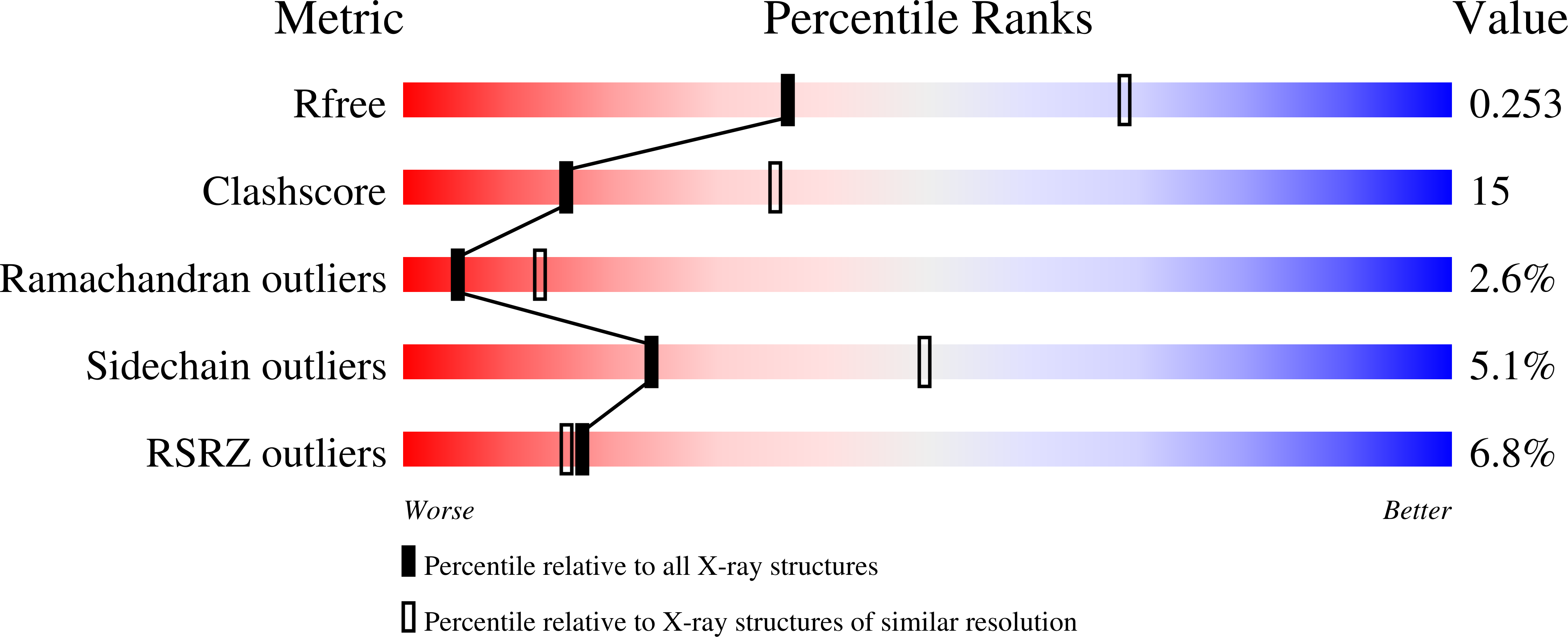
  • R-Value Free: 0.264 
  • R-Value Work: 0.218 
  • R-Value Observed: 0.218 

Starting Models: experimental
View more details

wwPDB Validation   3D Report Full Report


This is version 1.3 of the entry. See complete history


Literature

Specificity in Trk-Receptor:Neurotrophin Interaction: The Crystal Structure of Trkb-D5 in Complex with Neurotrophin-4/5

Banfield, M.J.Naylor, R.L.Robertson, A.G.S.Allen, S.J.Dawbarn, D.Brady, R.L.

(2001) Structure 9: 1191

  • DOI: https://doi.org/10.1016/s0969-2126(01)00681-5
  • Primary Citation of Related Structures:  
    1HCF

  • PubMed Abstract: 

    The binding of neurotrophin ligands to their respective Trk cellular receptors initiates intracellular signals essential for the growth and survival of neurons. The site of neurotrophin binding has been located to the fifth extracellular domain of the Trk receptor, with this region regulating both the affinity and specificity of Trk receptor:neurotrophin interaction. Neurotrophin function has been implicated in a number of neurological disorders, including Alzheimer's disease and Parkinson's disease. We have determined the 2.7 A crystal structure of neurotrophin-4/5 bound to the neurotrophin binding domain of its high-affinity receptor TrkB (TrkB-d5). As previously seen in the interaction of nerve growth factor with TrkA, neurotrophin-4/5 forms a crosslink between two spatially distant receptor molecules. The contacts formed in the TrkB-d5:neurotrophin-4/5 complex can be divided into a conserved area similar to a region observed in the TrkA-d5:NGF complex and a second site-unique in each ligand-receptor pair-formed primarily by the ordering of the neurotrophin N terminus. Together, the structures of the TrkB-d5:NT-4/5 and TrkA-d5:NGF complexes confirm a consistent pattern of recognition in Trk receptor:neurotrophin complex formation. In both cases, the N terminus of the neurotrophin becomes ordered only on complex formation. This ordering appears to be directed largely by the receptor surface, with the resulting complementary surfaces providing the main determinant of receptor specificity. These features provide an explanation both for the limited crossreactivity observed between the range of neurotrophins and Trk receptors and for the high-affinity binding associated with respective ligand-receptor pairs.


  • Organizational Affiliation

    Department of Biochemistry, University of Bristol, Bristol BS8 1TD, United Kingdom.


Macromolecules
Find similar proteins by:  (by identity cutoff)  |  3D Structure
Entity ID: 1
MoleculeChains Sequence LengthOrganismDetailsImage
NEUROTROPHIN-4
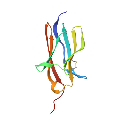
A, B
130Homo sapiensMutation(s): 0 
UniProt & NIH Common Fund Data Resources
Find proteins for P34130 (Homo sapiens)
Explore P34130 
Go to UniProtKB:  P34130
PHAROS:  P34130
GTEx:  ENSG00000225950 
Entity Groups  
Sequence Clusters30% Identity50% Identity70% Identity90% Identity95% Identity100% Identity
UniProt GroupP34130
Sequence Annotations
Expand
  • Reference Sequence
Find similar proteins by:  (by identity cutoff)  |  3D Structure
Entity ID: 2
MoleculeChains Sequence LengthOrganismDetailsImage
BDNF/NT-3 GROWTH FACTORS RECEPTORC [auth X],
D [auth Y]
101Homo sapiensMutation(s): 0 
EC: 2.7.10.1
UniProt & NIH Common Fund Data Resources
Find proteins for Q16620 (Homo sapiens)
Explore Q16620 
Go to UniProtKB:  Q16620
PHAROS:  Q16620
GTEx:  ENSG00000148053 
Entity Groups  
Sequence Clusters30% Identity50% Identity70% Identity90% Identity95% Identity100% Identity
UniProt GroupQ16620
Sequence Annotations
Expand
  • Reference Sequence
Experimental Data & Validation

Experimental Data

  • Method: X-RAY DIFFRACTION
  • Resolution: 2.70 Å
  • R-Value Free: 0.264 
  • R-Value Work: 0.218 
  • R-Value Observed: 0.218 
  • Space Group: P 21 21 21
Unit Cell:
Length ( Å )Angle ( ˚ )
a = 74.372α = 90
b = 80.406β = 90
c = 91.318γ = 90
Software Package:
Software NamePurpose
CNSrefinement
DENZOdata reduction
SCALEPACKdata scaling
AMoREphasing

Structure Validation

View Full Validation Report


Entry History 

Deposition Data

Revision History  (Full details and data files)

  • Version 1.0: 2001-12-06
    Type: Initial release
  • Version 1.1: 2011-05-08
    Changes: Version format compliance
  • Version 1.2: 2011-07-13
    Changes: Version format compliance
  • Version 1.3: 2023-12-13
    Changes: Data collection, Database references, Other, Refinement description