1FHI

SUBSTRATE ANALOG (IB2) COMPLEX WITH THE FRAGILE HISTIDINE TRIAD PROTEIN, FHIT


Experimental Data Snapshot

  • Method: X-RAY DIFFRACTION
  • Resolution: 3.10 Å
  • R-Value Free: 0.314 
  • R-Value Work: 0.241 
  • R-Value Observed: 0.241 

Starting Model: experimental
View more details

wwPDB Validation   3D Report Full Report


Ligand Structure Quality Assessment 


This is version 1.4 of the entry. See complete history


Literature

Genetic, biochemical, and crystallographic characterization of Fhit-substrate complexes as the active signaling form of Fhit.

Pace, H.C.Garrison, P.N.Robinson, A.K.Barnes, L.D.Draganescu, A.Rosler, A.Blackburn, G.M.Siprashvili, Z.Croce, C.M.Huebner, K.Brenner, C.

(1998) Proc Natl Acad Sci U S A 95: 5484-5489

  • DOI: https://doi.org/10.1073/pnas.95.10.5484
  • Primary Citation of Related Structures:  
    1FHI, 2FHI

  • PubMed Abstract: 

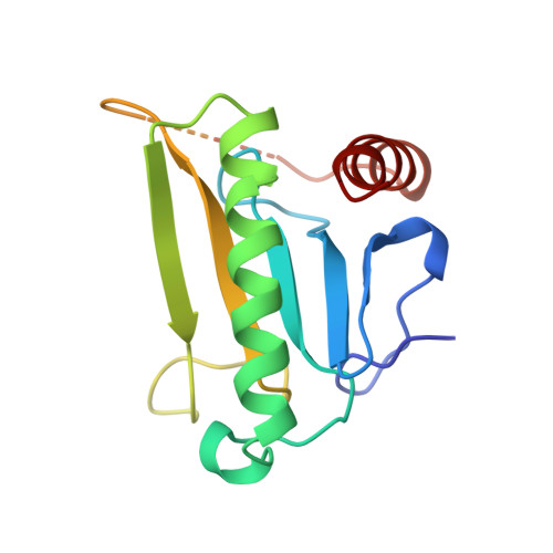
    Alterations in the FHIT gene at 3p14.2 occur as early and frequent events in the development of several common human cancers. The ability of human Fhit-negative cells to form tumors in nude mice is suppressed by stable reexpression of Fhit protein. Fhit protein is a diadenosine P1,P3-triphosphate (ApppA) hydrolase whose fungal and animal homologs form a branch of the histidine triad (HIT) superfamily of nucleotide-binding proteins. Because the His-96 --> Asn substitution of Fhit, which retards ApppA hydrolase activity by seven orders of magnitude, did not block tumor-suppressor activity in vivo, we determined whether this mutation affected ApppA binding or particular steps in the ApppA catalytic cycle. Evidence is presented that His-96 --> Asn protein binds ApppA well and forms an enzyme-AMP intermediate extremely poorly, suggesting that Fhit-substrate complexes are the likely signaling form of the enzyme. The cocrystal structure of Fhit bound to Ado-p-CH2-p-ps-Ado (IB2), a nonhydrolyzable ApppA analog, was refined to 3.1 A, and the structure of His-96 --> Asn Fhit with IB2 was refined to 2.6 A, revealing that two ApppA molecules bind per Fhit dimer; identifying two additional adenosine-binding sites on the dimer surface; and illustrating that His-98 is positioned to donate a hydrogen bond to the scissile bridging oxygen of ApppA substrates. The form of Fhit bound to two ApppA substrates would present to the cell a dramatically phosphorylated surface, prominently displaying six phosphate groups and two adenosine moieties in place of a deep cavity lined with histidines, arginines, and glutamines.


  • Organizational Affiliation

    Kimmel Cancer Institute, Thomas Jefferson University, Philadelphia, PA 19107, USA.


Macromolecules
Find similar proteins by:  (by identity cutoff)  |  3D Structure
Entity ID: 1
MoleculeChains Sequence LengthOrganismDetailsImage
FRAGILE HISTIDINE TRIAD PROTEIN147Homo sapiensMutation(s): 0 
Gene Names: FHIT
EC: 3.6.1.29
UniProt & NIH Common Fund Data Resources
Find proteins for P49789 (Homo sapiens)
Explore P49789 
Go to UniProtKB:  P49789
GTEx:  ENSG00000189283 
Entity Groups  
Sequence Clusters30% Identity50% Identity70% Identity90% Identity95% Identity100% Identity
UniProt GroupP49789
Sequence Annotations
Expand
  • Reference Sequence
Small Molecules
Ligands 1 Unique
IDChains Name / Formula / InChI Key2D Diagram3D Interactions
IB2
Query on IB2

Download Ideal Coordinates CCD File 
B [auth A]P1-P2-METHYLENE-P3-THIO-DIADENOSINE TRIPHOSPHATE
C21 H29 N10 O14 P3 S
UJCWOSLCGXVJOD-LCHUORCTSA-N
Experimental Data & Validation

Experimental Data

  • Method: X-RAY DIFFRACTION
  • Resolution: 3.10 Å
  • R-Value Free: 0.314 
  • R-Value Work: 0.241 
  • R-Value Observed: 0.241 
  • Space Group: P 61 2 2
Unit Cell:
Length ( Å )Angle ( ˚ )
a = 50.728α = 90
b = 50.728β = 90
c = 268.513γ = 120
Software Package:
Software NamePurpose
X-PLORmodel building
X-PLORrefinement
DENZOdata reduction
SCALEPACKdata scaling
X-PLORphasing

Structure Validation

View Full Validation Report


Ligand Structure Quality Assessment 


Entry History 

Deposition Data

Revision History  (Full details and data files)

  • Version 1.0: 1998-06-17
    Type: Initial release
  • Version 1.1: 2008-03-24
    Changes: Version format compliance
  • Version 1.2: 2011-07-13
    Changes: Version format compliance
  • Version 1.3: 2023-08-09
    Changes: Database references, Derived calculations, Other, Refinement description
  • Version 1.4: 2024-05-22
    Changes: Data collection