8XB3

Structural mechanism of substrate binding and inhibition of the human Norepinephrine Transporter


Experimental Data Snapshot

  • Method: ELECTRON MICROSCOPY
  • Resolution: 2.80 Å
  • Aggregation State: PARTICLE 
  • Reconstruction Method: SINGLE PARTICLE 

wwPDB Validation   3D Report Full Report


This is version 1.1 of the entry. See complete history


Literature

Substrate binding and inhibition mechanism of norepinephrine transporter.

Ji, W.Miao, A.Liang, K.Liu, J.Qi, Y.Zhou, Y.Duan, X.Sun, J.Lai, L.Wu, J.X.

(2024) Nature 

  • DOI: https://doi.org/10.1038/s41586-024-07810-5
  • Primary Citation of Related Structures:  
    8XB2, 8XB3, 8XB4

  • PubMed Abstract: 

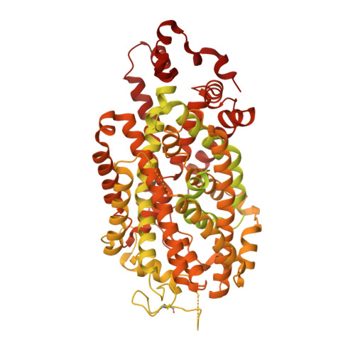
    Norepinephrine transporter (NET; encoded by SLC6A2) reuptakes the majority of the released noradrenaline back to the presynaptic terminals, thereby affecting the synaptic noradrenaline level 1 . Genetic mutations and dysregulation of NET are associated with a spectrum of neurological conditions in humans, making NET an important therapeutic target 1 . However, the structure and mechanism of NET remain unclear. Here we provide cryogenic electron microscopy structures of the human NET (hNET) in three functional states-the apo state, and in states bound to the substrate meta-iodobenzylguanidine (MIBG) or the orthosteric inhibitor radafaxine. These structures were captured in an inward-facing conformation, with a tightly sealed extracellular gate and an open intracellular gate. The substrate MIBG binds at the centre of hNET. Radafaxine also occupies the substrate-binding site and might block the structural transition of hNET for inhibition. These structures provide insights into the mechanism of substrate recognition and orthosteric inhibition of hNET.


  • Organizational Affiliation

    State Key Laboratory of Bioactive Substance and Function of Natural Medicines, Institute of Materia Medica, Chinese Academy of Medical Sciences & Peking Union Medical College, Beijing, P. R. China.


Macromolecules
Find similar proteins by:  (by identity cutoff)  |  3D Structure
Entity ID: 1
MoleculeChains Sequence LengthOrganismDetailsImage
GFP-MBP-solute carrier family 6 member 21,244Homo sapiensEscherichia coli K-12Mutation(s): 0 
Gene Names: SLC6A2malE
Membrane Entity: Yes 
UniProt & NIH Common Fund Data Resources
Find proteins for P0AEX9 (Escherichia coli (strain K12))
Explore P0AEX9 
Go to UniProtKB:  P0AEX9
Find proteins for P23975 (Homo sapiens)
Explore P23975 
Go to UniProtKB:  P23975
PHAROS:  P23975
GTEx:  ENSG00000103546 
Entity Groups  
Sequence Clusters30% Identity50% Identity70% Identity90% Identity95% Identity100% Identity
UniProt GroupsP23975P0AEX9
Glycosylation
Glycosylation Sites: 1Go to GlyGen: P23975-1
Sequence Annotations
Expand
  • Reference Sequence
Small Molecules
Ligands 2 Unique
IDChains Name / Formula / InChI Key2D Diagram3D Interactions
YMN (Subject of Investigation/LOI)
Query on YMN

Download Ideal Coordinates CCD File 
B [auth A]1-[(3-iodanylphenyl)methyl]guanidine
C8 H10 I N3
PDWUPXJEEYOOTR-UHFFFAOYSA-N
NAG
Query on NAG

Download Ideal Coordinates CCD File 
C [auth A]2-acetamido-2-deoxy-beta-D-glucopyranose
C8 H15 N O6
OVRNDRQMDRJTHS-FMDGEEDCSA-N
Experimental Data & Validation

Experimental Data

  • Method: ELECTRON MICROSCOPY
  • Resolution: 2.80 Å
  • Aggregation State: PARTICLE 
  • Reconstruction Method: SINGLE PARTICLE 
EM Software:
TaskSoftware PackageVersion
MODEL REFINEMENTPHENIX1.16_3549:

Structure Validation

View Full Validation Report


Entry History & Funding Information

Deposition Data


Funding OrganizationLocationGrant Number
National Natural Science Foundation of China (NSFC)China32371266

Revision History  (Full details and data files)

  • Version 1.0: 2024-06-26
    Type: Initial release
  • Version 1.1: 2024-08-28
    Changes: Data collection, Database references