


Substrate-bound crystal structure of a P450 enzyme DmlH that catalyze intramolecular phenol coupling in the biosynthesis of cihanmycins
Fang, C., Zhang, L., Zhu, Y., Zhang, C.To be published.
Experimental Data Snapshot
Starting Model: experimental
View more details
Entity ID: 1 | |||||
|---|---|---|---|---|---|
| Molecule | Chains | Sequence Length | Organism | Details | Image |
| Cytochrome P450 | 428 | Streptomyces sp. | Mutation(s): 0 |  | |
Entity Groups | |||||
| Sequence Clusters | 30% Identity50% Identity70% Identity90% Identity95% Identity100% Identity | ||||
Sequence AnnotationsExpand | |||||
| |||||
Find similar proteins by: Sequence | 3D Structure
Entity ID: 2 | |||||
|---|---|---|---|---|---|
| Molecule | Chains | Sequence Length | Organism | Details | Image |
| THR-D4P-GLU-GLY-BB8-PRO-KJW-GLY-A1D5P-DPN | 10 | Streptomyces sp. | Mutation(s): 0 | 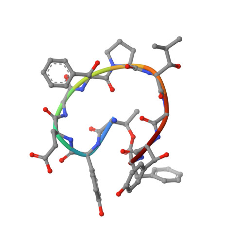 | |
Entity Groups | |||||
| Sequence Clusters | 30% Identity50% Identity70% Identity90% Identity95% Identity100% Identity | ||||
Sequence AnnotationsExpand | |||||
| |||||
| Ligands 2 Unique | |||||
|---|---|---|---|---|---|
| ID | Chains | Name / Formula / InChI Key | 2D Diagram | 3D Interactions | |
| HEM Query on HEM | C [auth A] | PROTOPORPHYRIN IX CONTAINING FE C34 H32 Fe N4 O4 KABFMIBPWCXCRK-RGGAHWMASA-L |  | ||
| R8L (Subject of Investigation/LOI) Query on R8L | D [auth B] | (E)-3-[2-[(2R,3S)-3-[(1R)-1-aminocarbonyloxypropyl]oxiran-2-yl]phenyl]prop-2-enoic acid C15 H17 N O5 APIHDLCQFHISLU-PQPVHHHVSA-N |  | ||
| Modified Residues 2 Unique | |||||
|---|---|---|---|---|---|
| ID | Chains | Type | Formula | 2D Diagram | Parent |
| A1D5P Query on A1D5P | B | D-PEPTIDE LINKING | C9 H11 N O4 |  | TYR |
| BB8 Query on BB8 | B | L-PEPTIDE LINKING | C9 H11 N O3 |  | PHE |
Entity ID: 2 | |||||
|---|---|---|---|---|---|
| ID | Chains | Name | Type/Class | 2D Diagram | 3D Interactions |
| PRD_002429 Query on PRD_002429 | B | cihanmycin G | Cyclic peptide / Unknown |  | |
| Length ( Å ) | Angle ( ˚ ) |
|---|---|
| a = 36.082 | α = 90 |
| b = 100.162 | β = 90 |
| c = 105.811 | γ = 90 |
| Software Name | Purpose |
|---|---|
| PHENIX | refinement |
| Aimless | data scaling |
| MOLREP | phasing |
| CrysalisPro | data reduction |
| Funding Organization | Location | Grant Number |
|---|---|---|
| National Natural Science Foundation of China (NSFC) | China | 22177118 |
| National Natural Science Foundation of China (NSFC) | China | 31820103003 |
| National Natural Science Foundation of China (NSFC) | China | 31630004 |
RCSB PDB (citation) is hosted by
RCSB PDB is a member of the

