8VM2

Crystal Structure of NRAS Q61K bound to GTP


Experimental Data Snapshot

  • Method: X-RAY DIFFRACTION
  • Resolution: 1.74 Å
  • R-Value Free: 0.203 
  • R-Value Work: 0.172 
  • R-Value Observed: 0.173 

Starting Model: experimental
View more details

wwPDB Validation   3D Report Full Report


Ligand Structure Quality Assessment 


This is version 1.0 of the entry. See complete history


Literature

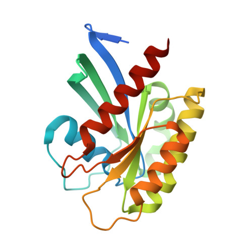
Crystal structure of NRAS Q61K with a ligand-induced pocket near switch II.

Gebregiworgis, T.Chan, J.Y.Kuntz, D.A.Prive, G.G.Marshall, C.B.Ikura, M.

(2024) Eur J Cell Biol 103: 151414-151414

  • DOI: https://doi.org/10.1016/j.ejcb.2024.151414
  • Primary Citation of Related Structures:  
    8VM2

  • PubMed Abstract: 

    The RAS isoforms (KRAS, HRAS and NRAS) have distinct cancer type-specific profiles. NRAS mutations are the second most prevalent RAS mutations in skin and hematological malignancies. Although RAS proteins were considered undruggable for decades, isoform and mutation-specific investigations have produced successful RAS inhibitors that are either specific to certain mutants, isoforms (pan-KRAS) or target all RAS proteins (pan-RAS). While extensive structural and biochemical investigations have focused mainly on K- and H-RAS mutations, NRAS mutations have received less attention, and the most prevalent NRAS mutations in human cancers, Q61K and Q61R, are rare in K- and H-RAS. This manuscript presents a crystal structure of the NRAS Q61K mutant in the GTP-bound form. Our structure reveals a previously unseen pocket near switch II induced by the binding of a ligand to the active form of the protein. This observation reveals a binding site that can potentially be exploited for development of inhibitors against mutant NRAS. Furthermore, the well-resolved catalytic site of this GTPase bound to native GTP provides insight into the stalled GTP hydrolysis observed for NRAS-Q61K.


  • Organizational Affiliation

    Princess Margaret Cancer Centre, University Health Network, Toronto, Ontario M5G 1L7, Canada; Department of Biochemistry, Schulich School of Medicine and Dentistry, Western University, London, Ontario N6A 5C1, Canada; Department of Oncology, Schulich School of Medicine and Dentistry, Western University, London, Ontario N6A 5W9, Canada. Electronic address: tgebregi@uwo.ca.


Macromolecules
Find similar proteins by:  (by identity cutoff)  |  3D Structure
Entity ID: 1
MoleculeChains Sequence LengthOrganismDetailsImage
GTPase NRas
A, B, C
190Homo sapiensMutation(s): 1 
Gene Names: NRASHRAS1
EC: 3.6.5.2
UniProt & NIH Common Fund Data Resources
Find proteins for P01111 (Homo sapiens)
Explore P01111 
Go to UniProtKB:  P01111
PHAROS:  P01111
GTEx:  ENSG00000213281 
Entity Groups  
Sequence Clusters30% Identity50% Identity70% Identity90% Identity95% Identity100% Identity
UniProt GroupP01111
Sequence Annotations
Expand
  • Reference Sequence
Small Molecules
Ligands 3 Unique
IDChains Name / Formula / InChI Key2D Diagram3D Interactions
GTP (Subject of Investigation/LOI)
Query on GTP

Download Ideal Coordinates CCD File 
D [auth A],
F [auth A],
H [auth B],
J [auth B],
K [auth C]
GUANOSINE-5'-TRIPHOSPHATE
C10 H16 N5 O14 P3
XKMLYUALXHKNFT-UUOKFMHZSA-N
MG
Query on MG

Download Ideal Coordinates CCD File 
E [auth A],
I [auth B],
L [auth C]
MAGNESIUM ION
Mg
JLVVSXFLKOJNIY-UHFFFAOYSA-N
UNX
Query on UNX

Download Ideal Coordinates CCD File 
G [auth A],
M [auth C]
UNKNOWN ATOM OR ION
X
Experimental Data & Validation

Experimental Data

Unit Cell:
Length ( Å )Angle ( ˚ )
a = 75.789α = 90
b = 55.699β = 98.15
c = 149.291γ = 90
Software Package:
Software NamePurpose
PHENIXrefinement
CrysalisProdata collection
CrysalisProdata reduction
Aimlessdata scaling
PHASERphasing

Structure Validation

View Full Validation Report


Ligand Structure Quality Assessment 


Entry History & Funding Information

Deposition Data


Funding OrganizationLocationGrant Number
Canadian Institutes of Health Research (CIHR)CanadaFDN-1542284

Revision History  (Full details and data files)

  • Version 1.0: 2024-05-01
    Type: Initial release