7RNT

CRYSTAL STRUCTURE OF THE TYR45TRP MUTANT OF RIBONUCLEASE T1 IN A COMPLEX WITH 2'-ADENYLIC ACID


Experimental Data Snapshot

  • Method: X-RAY DIFFRACTION
  • Resolution: 1.90 Å
  • R-Value Observed: 0.170 

wwPDB Validation   3D Report Full Report


This is version 1.3 of the entry. See complete history


Literature

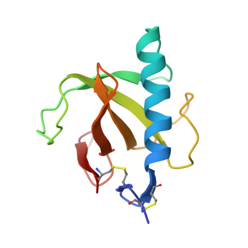
Crystal structure of the Tyr45Trp mutant of ribonuclease T1 in a complex with 2'-adenylic acid.

Koellner, G.Grunert, H.P.Landt, O.Saenger, W.

(1991) Eur J Biochem 201: 199-202

  • DOI: https://doi.org/10.1111/j.1432-1033.1991.tb16274.x
  • Primary Citation of Related Structures:  
    7RNT

  • PubMed Abstract: 

    The recombinant Tyr45Trp mutant of Lys25-ribonuclease T1 was overexpressed and purified from an Escherichia coli strain. The mutant enzyme, which shows reduced activity towards GpA and increased activity towards pGpC, pApC and pUpC compared with wild-type RNase T1, was co-crystallized with 2'-adenylic acid by microdialysis. The space group is P212121 with unit cell dimenions a = 4.932(2), b = 4.661(2), c = 4.092(1) nm. The crystal structure was solved using the coordinates of the isomorphous complex of wild-type RNase T1 with 2'-AMP. The refinement was based on Fhkl of 7726 reflexions with Fo greater than or equal to 1 sigma (Fo) in the resolution range of 2.0-0.19 nm and converged with an R factor of 0.179. The adenosine of 2'-AMP is not bound to the guanosine binding site, as could be expected from the mutation of Tyr45Trp, but is stacked on the Gly74 carbonyl group and the His92 imidazole group which form a subsite for substrate binding, as already observed in the wild-type 2'-AMP complex. The point mutation of Tyr45Trp does not perturb the backbone conformation and the Trp-indole side chain is in a comparable position to the phenolic Tyr45 of the wild-type enzyme.


  • Organizational Affiliation

    Institut für Kristallographie, Freie Universität Berlin, Federal Republic of Germany.


Macromolecules
Find similar proteins by:  (by identity cutoff)  |  3D Structure
Entity ID: 1
MoleculeChains Sequence LengthOrganismDetailsImage
RIBONUCLEASE T1104Aspergillus oryzaeMutation(s): 0 
EC: 3.1.27.3
UniProt
Find proteins for P00651 (Aspergillus oryzae (strain ATCC 42149 / RIB 40))
Explore P00651 
Go to UniProtKB:  P00651
Entity Groups  
Sequence Clusters30% Identity50% Identity70% Identity90% Identity95% Identity100% Identity
UniProt GroupP00651
Sequence Annotations
Expand
  • Reference Sequence
Small Molecules
Ligands 2 Unique
IDChains Name / Formula / InChI Key2D Diagram3D Interactions
2AM
Query on 2AM

Download Ideal Coordinates CCD File 
C [auth A]ADENOSINE-2'-MONOPHOSPHATE
C10 H14 N5 O7 P
QDFHPFSBQFLLSW-KQYNXXCUSA-N
CA
Query on CA

Download Ideal Coordinates CCD File 
B [auth A]CALCIUM ION
Ca
BHPQYMZQTOCNFJ-UHFFFAOYSA-N
Experimental Data & Validation

Experimental Data

  • Method: X-RAY DIFFRACTION
  • Resolution: 1.90 Å
  • R-Value Observed: 0.170 
  • Space Group: P 21 21 21
Unit Cell:
Length ( Å )Angle ( ˚ )
a = 49.32α = 90
b = 46.61β = 90
c = 40.92γ = 90
Software Package:
Software NamePurpose
TNTrefinement

Structure Validation

View Full Validation Report


Entry History 

Deposition Data

Revision History  (Full details and data files)

  • Version 1.0: 1993-01-15
    Type: Initial release
  • Version 1.1: 2008-03-25
    Changes: Version format compliance
  • Version 1.2: 2011-07-13
    Changes: Version format compliance
  • Version 1.3: 2017-11-29
    Changes: Derived calculations, Other