7PME

Cryo-EM structure of the actomyosin-V complex in the post-rigor transition state (AppNHp, central 3er/2er)


Experimental Data Snapshot

  • Method: ELECTRON MICROSCOPY
  • Resolution: 2.90 Å
  • Aggregation State: FILAMENT 
  • Reconstruction Method: HELICAL 

wwPDB Validation   3D Report Full Report


This is version 1.0 of the entry. See complete history


Literature

High-resolution structures of the actomyosin-V complex in three nucleotide states provide insights into the force generation mechanism.

Pospich, S.Sweeney, H.L.Houdusse, A.Raunser, S.

(2021) Elife 10

  • DOI: https://doi.org/10.7554/eLife.73724
  • Primary Citation of Related Structures:  
    7PLT, 7PLU, 7PLV, 7PLW, 7PLX, 7PLY, 7PLZ, 7PM0, 7PM1, 7PM2, 7PM3, 7PM5, 7PM6, 7PM7, 7PM8, 7PM9, 7PMA, 7PMB, 7PMC, 7PMD, 7PME, 7PMF, 7PMG, 7PMH, 7PMI, 7PMJ, 7PML

  • PubMed Abstract: 

    The molecular motor myosin undergoes a series of major structural transitions during its force-producing motor cycle. The underlying mechanism and its coupling to ATP hydrolysis and actin binding are only partially understood, mostly due to sparse structural data on actin-bound states of myosin. Here, we report 26 high-resolution cryo-EM structures of the actomyosin-V complex in the strong-ADP, rigor, and a previously unseen post-rigor transition state that binds the ATP analog AppNHp. The structures reveal a high flexibility of myosin in each state and provide valuable insights into the structural transitions of myosin-V upon ADP release and binding of AppNHp, as well as the actomyosin interface. In addition, they show how myosin is able to specifically alter the structure of F-actin.


  • Organizational Affiliation

    Department of Structural Biochemistry, Max Planck Institute of Molecular Physiology, Dortmund, Germany.


Macromolecules
Find similar proteins by:  (by identity cutoff)  |  3D Structure
Entity ID: 1
MoleculeChains Sequence LengthOrganismDetailsImage
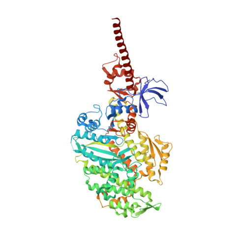
Unconventional myosin-VaA,
E [auth D]
792Gallus gallusMutation(s): 0 
Gene Names: MYO5A
UniProt
Find proteins for Q02440 (Gallus gallus)
Explore Q02440 
Go to UniProtKB:  Q02440
Entity Groups  
Sequence Clusters30% Identity50% Identity70% Identity90% Identity95% Identity100% Identity
UniProt GroupQ02440
Sequence Annotations
Expand
  • Reference Sequence
Find similar proteins by:  (by identity cutoff)  |  3D Structure
Entity ID: 2
MoleculeChains Sequence LengthOrganismDetailsImage
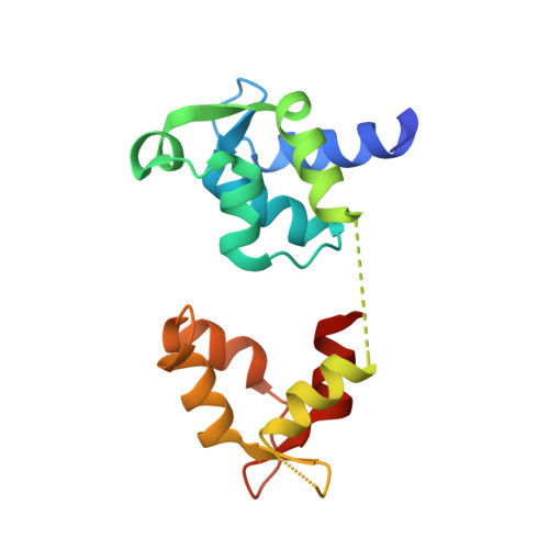
Myosin light chain 6BB,
F [auth E]
151Homo sapiensMutation(s): 0 
Gene Names: MYL6BMLC1SA
UniProt & NIH Common Fund Data Resources
Find proteins for P14649 (Homo sapiens)
Explore P14649 
Go to UniProtKB:  P14649
PHAROS:  P14649
GTEx:  ENSG00000196465 
Entity Groups  
Sequence Clusters30% Identity50% Identity70% Identity90% Identity95% Identity100% Identity
UniProt GroupP14649
Sequence Annotations
Expand
  • Reference Sequence
Find similar proteins by:  (by identity cutoff)  |  3D Structure
Entity ID: 3
MoleculeChains Sequence LengthOrganismDetailsImage
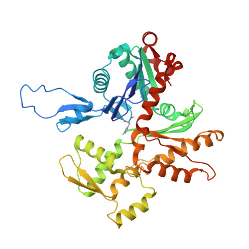
Actin, alpha skeletal muscleC,
G [auth F],
I [auth G]
377Oryctolagus cuniculusMutation(s): 0 
EC: 3.6.4
UniProt
Find proteins for P68135 (Oryctolagus cuniculus)
Explore P68135 
Go to UniProtKB:  P68135
Entity Groups  
Sequence Clusters30% Identity50% Identity70% Identity90% Identity95% Identity100% Identity
UniProt GroupP68135
Sequence Annotations
Expand
  • Reference Sequence

Find similar proteins by:  Sequence   |   3D Structure  

Entity ID: 4
MoleculeChains Sequence LengthOrganismDetailsImage
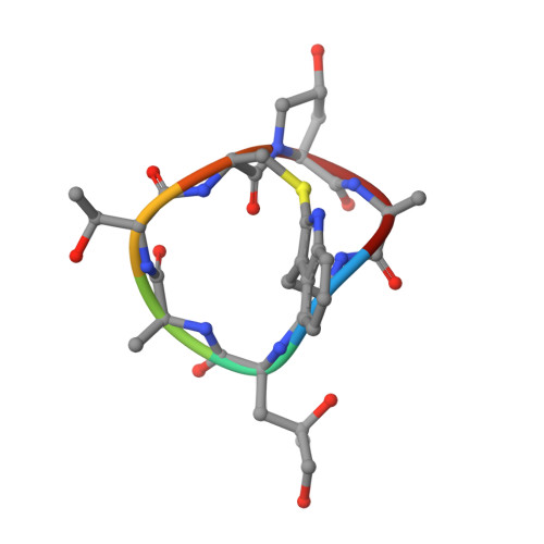
PhalloidinD [auth H],
H [auth I],
J
7Amanita phalloidesMutation(s): 0 
Sequence Annotations
Expand
  • Reference Sequence
Small Molecules
Ligands 3 Unique
IDChains Name / Formula / InChI Key2D Diagram3D Interactions
ANP
Query on ANP

Download Ideal Coordinates CCD File 
K [auth A],
P [auth D]
PHOSPHOAMINOPHOSPHONIC ACID-ADENYLATE ESTER
C10 H17 N6 O12 P3
PVKSNHVPLWYQGJ-KQYNXXCUSA-N
ADP
Query on ADP

Download Ideal Coordinates CCD File 
N [auth C],
S [auth F],
U [auth G]
ADENOSINE-5'-DIPHOSPHATE
C10 H15 N5 O10 P2
XTWYTFMLZFPYCI-KQYNXXCUSA-N
MG
Query on MG

Download Ideal Coordinates CCD File 
L [auth A]
M [auth A]
O [auth C]
Q [auth D]
R [auth D]
L [auth A],
M [auth A],
O [auth C],
Q [auth D],
R [auth D],
T [auth F],
V [auth G]
MAGNESIUM ION
Mg
JLVVSXFLKOJNIY-UHFFFAOYSA-N
Modified Residues  2 Unique
IDChains TypeFormula2D DiagramParent
HIC
Query on HIC
C,
G [auth F],
I [auth G]
L-PEPTIDE LINKINGC7 H11 N3 O2HIS
HYP
Query on HYP
D [auth H],
H [auth I],
J
L-PEPTIDE LINKINGC5 H9 N O3PRO
Biologically Interesting Molecules (External Reference) 1 Unique
Experimental Data & Validation

Experimental Data

  • Method: ELECTRON MICROSCOPY
  • Resolution: 2.90 Å
  • Aggregation State: FILAMENT 
  • Reconstruction Method: HELICAL 
EM Software:
TaskSoftware PackageVersion
RECONSTRUCTIONSPHIRE
MODEL REFINEMENTPHENIX1.17
MODEL REFINEMENTISOLDE0.91

Structure Validation

View Full Validation Report


Entry History & Funding Information

Deposition Data


Funding OrganizationLocationGrant Number
Max Planck SocietyGermany--
European CommissionEuropean UnionERC-2019-SyG 856118
Centre National de la Recherche Scientifique (CNRS)France--
Agence Nationale de la Recherche (ANR)FranceANR-17-CE11-0029-01
National Institutes of Health/National Institute on Deafness and Other Communication Disorders (NIH/NIDCD)United StatesR01-DC009100

Revision History  (Full details and data files)

  • Version 1.0: 2021-12-22
    Type: Initial release