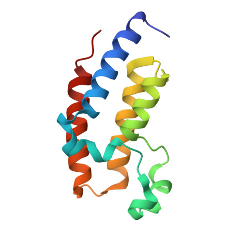
7EO5

Crystal structure of pyrano 1,3, oxazine derivative in complex with the second bromodomain of BRD2


Experimental Data Snapshot

  • Method: X-RAY DIFFRACTION
  • Resolution: 2.00 Å
  • R-Value Free: 0.224 
  • R-Value Work: 0.174 
  • R-Value Observed: 0.176 

Starting Model: experimental
View more details

wwPDB Validation   3D Report Full Report


Ligand Structure Quality Assessment 


This is version 1.2 of the entry. See complete history


Literature

Structural investigation of a pyrano-1,3-oxazine derivative and the phenanthridinone core moiety against BRD2 bromodomains.

Arole, A.H.Deshmukh, P.Sridhar, A.Padmanabhan, B.

(2022) Acta Crystallogr F Struct Biol Commun 78: 119-127

  • DOI: https://doi.org/10.1107/S2053230X22001066
  • Primary Citation of Related Structures:  
    7ENV, 7ENZ, 7EO5

  • PubMed Abstract: 

    The BET (bromodomain and extra-terminal) family of proteins recognize the acetylated histone code on chromatin and play important roles in transcriptional co-regulation. BRD2 and BRD4, which belong to the BET family, are promising drug targets for the management of chronic diseases. The discovery of new scaffold molecules, a pyrano-1,3-oxazine derivative (NSC 328111; NS5) and phenanthridinone-based derivatives (L10 and its core moiety L10a), as inhibitors of BRD2 bromodomains BD1 and BD2, respectively, has recently been reported. The compound NS5 has a significant inhibitory effect on BRD2 in glioblastoma. Here, the crystal structure of BRD2 BD2 in complex with NS5, refined to 2.0 Å resolution, is reported. Moreover, as the previously reported crystal structures of the BD1-NS5 complex and the BD2-L10a complex possess moderate electron density corresponding to the respective ligands, the crystal structures of these complexes were re-evaluated using new X-ray data. Together with biochemical studies using wild-type BRD2 BD1 and BD2 and various mutants, it is confirmed that the pyrano-1,3-oxazine and phenanthridinone derivatives are indeed potent inhibitors of BRD2 bromodomains.


  • Organizational Affiliation

    Department of Biophysics, National Institute of Mental Health and Neuro Sciences (NIMHANS), Hosur Main Road, Bengaluru 560 029, India.


Macromolecules
Find similar proteins by:  (by identity cutoff)  |  3D Structure
Entity ID: 1
MoleculeChains Sequence LengthOrganismDetailsImage
Bromodomain-containing protein 2115Homo sapiensMutation(s): 0 
Gene Names: BRD2KIAA9001RING3
UniProt & NIH Common Fund Data Resources
Find proteins for P25440 (Homo sapiens)
Explore P25440 
Go to UniProtKB:  P25440
PHAROS:  P25440
GTEx:  ENSG00000204256 
Entity Groups  
Sequence Clusters30% Identity50% Identity70% Identity90% Identity95% Identity100% Identity
UniProt GroupP25440
Sequence Annotations
Expand
  • Reference Sequence
Small Molecules
Ligands 2 Unique
IDChains Name / Formula / InChI Key2D Diagram3D Interactions
BUX (Subject of Investigation/LOI)
Query on BUX

Download Ideal Coordinates CCD File 
C [auth A]7-chloranyl-2-[(3-chlorophenyl)amino]pyrano[3,4-e][1,3]oxazine-4,5-dione
C13 H6 Cl2 N2 O4
YBBCXMKGFBMRLJ-UHFFFAOYSA-N
PGE
Query on PGE

Download Ideal Coordinates CCD File 
B [auth A]TRIETHYLENE GLYCOL
C6 H14 O4
ZIBGPFATKBEMQZ-UHFFFAOYSA-N
Experimental Data & Validation

Experimental Data

  • Method: X-RAY DIFFRACTION
  • Resolution: 2.00 Å
  • R-Value Free: 0.224 
  • R-Value Work: 0.174 
  • R-Value Observed: 0.176 
  • Space Group: P 21 21 2
Unit Cell:
Length ( Å )Angle ( ˚ )
a = 52.102α = 90
b = 71.121β = 90
c = 31.852γ = 90
Software Package:
Software NamePurpose
PHENIXrefinement
HKL-3000data reduction
HKL-3000data scaling
PHENIXphasing

Structure Validation

View Full Validation Report


Ligand Structure Quality Assessment 


Entry History 

Deposition Data

Revision History  (Full details and data files)

  • Version 1.0: 2022-03-09
    Type: Initial release
  • Version 1.1: 2022-03-16
    Changes: Database references
  • Version 1.2: 2023-11-29
    Changes: Data collection, Refinement description