


The crystal structure of chloramphenicol acetyltransferase-like protein from Vibrio fischeri ES114 in complex with taurocholic acid
Tan, K., Maltseva, N., Jedrzejczak, R., Kuhn, M., Joachimiak, A.To be published.
Experimental Data Snapshot
Starting Model: experimental
View more details
Entity ID: 1 | |||||
|---|---|---|---|---|---|
| Molecule | Chains | Sequence Length | Organism | Details | Image |
| Chloramphenicol acetyltransferase | 221 | Aliivibrio fischeri ES114 | Mutation(s): 0 Gene Names: VF_A0790 EC: 2.3.1.28 | 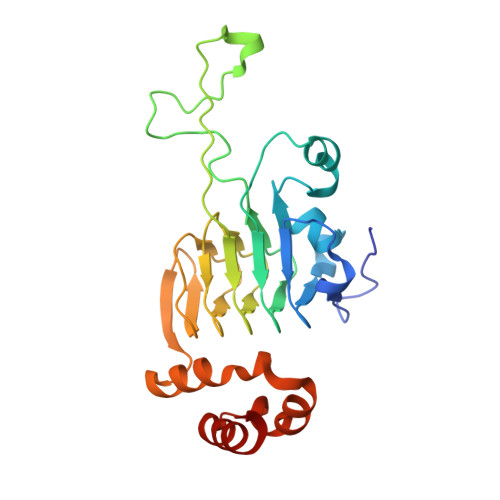 | |
UniProt | |||||
Find proteins for Q5DZD6 (Aliivibrio fischeri (strain ATCC 700601 / ES114)) Explore Q5DZD6 Go to UniProtKB: Q5DZD6 | |||||
Entity Groups | |||||
| Sequence Clusters | 30% Identity50% Identity70% Identity90% Identity95% Identity100% Identity | ||||
| UniProt Group | Q5DZD6 | ||||
Sequence AnnotationsExpand | |||||
| |||||
| Ligands 6 Unique | |||||
|---|---|---|---|---|---|
| ID | Chains | Name / Formula / InChI Key | 2D Diagram | 3D Interactions | |
| TCH (Subject of Investigation/LOI) Query on TCH | AA [auth E] BB [auth K] DA [auth F] GB [auth L] HA [auth G] | TAUROCHOLIC ACID C26 H45 N O7 S WBWWGRHZICKQGZ-HZAMXZRMSA-N |  | ||
| SO4 Query on SO4 | LA [auth G], O [auth A], VA [auth J], WA [auth J] | SULFATE ION O4 S QAOWNCQODCNURD-UHFFFAOYSA-L |  | ||
| GOL Query on GOL | CA [auth E] GA [auth F] OA [auth H] RA [auth I] T [auth B] | GLYCEROL C3 H8 O3 PEDCQBHIVMGVHV-UHFFFAOYSA-N |  | ||
| ACT Query on ACT | MA [auth G], P [auth A], Q [auth A] | ACETATE ION C2 H3 O2 QTBSBXVTEAMEQO-UHFFFAOYSA-M |  | ||
| FMT Query on FMT | AB [auth J] DB [auth K] EB [auth K] FB [auth K] XA [auth J] | FORMIC ACID C H2 O2 BDAGIHXWWSANSR-UHFFFAOYSA-N |  | ||
| CL Query on CL | BA [auth E] CB [auth K] EA [auth F] FA [auth F] HB [auth L] | CHLORIDE ION Cl VEXZGXHMUGYJMC-UHFFFAOYSA-M |  | ||
| Length ( Å ) | Angle ( ˚ ) |
|---|---|
| a = 43.519 | α = 89.399 |
| b = 121.363 | β = 89.911 |
| c = 146.17 | γ = 87.602 |
| Software Name | Purpose |
|---|---|
| SBC-Collect | data collection |
| PHENIX | refinement |
| HKL-3000 | data reduction |
| HKL-3000 | data scaling |
| HKL-3000 | phasing |
| Funding Organization | Location | Grant Number |
|---|---|---|
| National Institutes of Health/National Institute Of Allergy and Infectious Diseases (NIH/NIAID) | United States | HHSN272201700060C |
RCSB PDB (citation) is hosted by
RCSB PDB is a member of the

