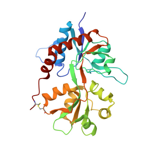
6F28

Crystal structure of the kainate receptor GluK3 ligand binding domain in complex with (S)-1-[2'-Amino-2'-carboxyethyl]-6-methyl-5,7-dihydropyrrolo[3,4-d]pyrimidin-2,4(1H,3H)-dione at resolution 2.4A


Experimental Data Snapshot

  • Method: X-RAY DIFFRACTION
  • Resolution: 2.40 Å
  • R-Value Free: 0.222 
  • R-Value Work: 0.195 
  • R-Value Observed: 0.197 

Starting Model: experimental
View more details

wwPDB Validation   3D Report Full Report


Ligand Structure Quality Assessment 


This is version 1.3 of the entry. See complete history


Literature

( S)-2-Amino-3-(5-methyl-3-hydroxyisoxazol-4-yl)propanoic Acid (AMPA) and Kainate Receptor Ligands: Further Exploration of Bioisosteric Replacements and Structural and Biological Investigation.

Brogi, S.Brindisi, M.Butini, S.Kshirsagar, G.U.Maramai, S.Chemi, G.Gemma, S.Campiani, G.Novellino, E.Fiorenzani, P.Pinassi, J.Aloisi, A.M.Gynther, M.Venskutonyte, R.Han, L.Frydenvang, K.Kastrup, J.S.Pickering, D.S.

(2018) J Med Chem 61: 2124-2130

  • DOI: https://doi.org/10.1021/acs.jmedchem.8b00099
  • Primary Citation of Related Structures:  
    6F28, 6F29

  • PubMed Abstract: 

    Starting from 1-4 and 7 structural templates, analogues based on bioisosteric replacements (5a-c vs 1, 2 and 6 vs 7) were synthesized for completing the SAR analysis. Interesting binding properties at GluA2, GluK1, and GluK3 receptors were discovered. The requirements for GluK3 interaction were elucidated by determining the X-ray structures of the GluK3-LBD with 2 and 5c and by computational studies. Antinociceptive potential was demonstrated for GluK1 partial agonist 3 and antagonist 7 (2 mg/kg ip).


  • Organizational Affiliation

    Department of Biotechnology, Chemistry and Pharmacy, (DoE 2018-2022) NatSynDrugs , University of Siena , Via A. Moro 2 , 53100 Siena , Italy.


Macromolecules
Find similar proteins by:  (by identity cutoff)  |  3D Structure
Entity ID: 1
MoleculeChains Sequence LengthOrganismDetailsImage
Glutamate receptor ionotropic, kainate 3,Glutamate receptor ionotropic, kainate 3
A, B
258Rattus norvegicusMutation(s): 0 
Gene Names: Grik3Glur7
UniProt
Find proteins for P42264 (Rattus norvegicus)
Explore P42264 
Go to UniProtKB:  P42264
Entity Groups  
Sequence Clusters30% Identity50% Identity70% Identity90% Identity95% Identity100% Identity
UniProt GroupP42264
Sequence Annotations
Expand
  • Reference Sequence
Small Molecules
Ligands 4 Unique
IDChains Name / Formula / InChI Key2D Diagram3D Interactions
CG8
Query on CG8

Download Ideal Coordinates CCD File 
D [auth A],
K [auth B]
(2~{S})-2-azanyl-3-[6-methyl-2,4-bis(oxidanylidene)-5,7-dihydropyrrolo[3,4-d]pyrimidin-1-yl]propanoic acid
C10 H14 N4 O4
BJXZWHLUHLDGNK-LURJTMIESA-N
SO4
Query on SO4

Download Ideal Coordinates CCD File 
L [auth B]SULFATE ION
O4 S
QAOWNCQODCNURD-UHFFFAOYSA-L
ZN
Query on ZN

Download Ideal Coordinates CCD File 
C [auth A],
G [auth B],
H [auth B],
I [auth B],
J [auth B]
ZINC ION
Zn
PTFCDOFLOPIGGS-UHFFFAOYSA-N
CL
Query on CL

Download Ideal Coordinates CCD File 
E [auth A],
F [auth A],
M [auth B]
CHLORIDE ION
Cl
VEXZGXHMUGYJMC-UHFFFAOYSA-M
Experimental Data & Validation

Experimental Data

  • Method: X-RAY DIFFRACTION
  • Resolution: 2.40 Å
  • R-Value Free: 0.222 
  • R-Value Work: 0.195 
  • R-Value Observed: 0.197 
  • Space Group: P 2 2 21
Unit Cell:
Length ( Å )Angle ( ˚ )
a = 131.039α = 90
b = 56.214β = 90
c = 87.189γ = 90
Software Package:
Software NamePurpose
SCALAdata scaling
PHENIXrefinement
PDB_EXTRACTdata extraction
XDSdata reduction
PHASERphasing

Structure Validation

View Full Validation Report


Ligand Structure Quality Assessment 


Entry History 

Deposition Data

Revision History  (Full details and data files)

  • Version 1.0: 2018-02-28
    Type: Initial release
  • Version 1.1: 2018-03-07
    Changes: Database references
  • Version 1.2: 2018-03-21
    Changes: Database references
  • Version 1.3: 2024-01-17
    Changes: Data collection, Database references, Derived calculations, Refinement description