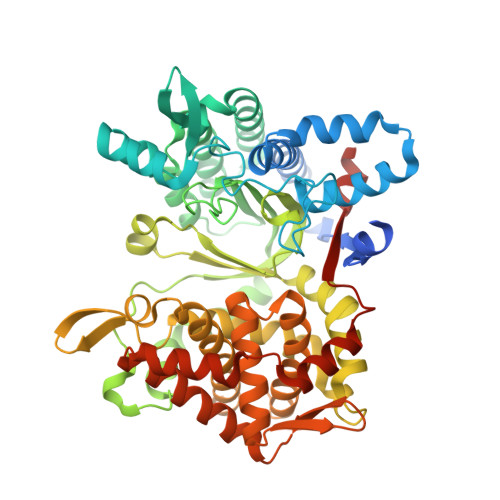
6EOM

Structure of DPP III from Caldithrix abyssi


Experimental Data Snapshot

  • Method: X-RAY DIFFRACTION
  • Resolution: 2.10 Å
  • R-Value Free: 0.240 
  • R-Value Work: 0.210 
  • R-Value Observed: 0.211 

Starting Model: experimental
View more details

wwPDB Validation   3D Report Full Report


This is version 1.1 of the entry. See complete history


Literature

The first dipeptidyl peptidase III from a thermophile: Structural basis for thermal stability and reduced activity.

Sabljic, I.Tomin, M.Matovina, M.Sucec, I.Tomasic Paic, A.Tomic, A.Abramic, M.Tomic, S.

(2018) PLoS One 13: e0192488-e0192488

  • DOI: https://doi.org/10.1371/journal.pone.0192488
  • Primary Citation of Related Structures:  
    6EOM

  • PubMed Abstract: 

    Dipeptidyl peptidase III (DPP III) isolated from the thermophilic bacteria Caldithrix abyssi (Ca) is a two-domain zinc exopeptidase, a member of the M49 family. Like other DPPs III, it cleaves dipeptides from the N-terminus of its substrates but differently from human, yeast and Bacteroides thetaiotaomicron (mesophile) orthologs, it has the pentapeptide zinc binding motif (HEISH) in the active site instead of the hexapeptide (HEXXGH). The aim of our study was to investigate structure, dynamics and activity of CaDPP III, as well as to find possible differences with already characterized DPPs III from mesophiles, especially B. thetaiotaomicron. The enzyme structure was determined by X-ray diffraction, while stability and flexibility were investigated using MD simulations. Using molecular modeling approach we determined the way of ligands binding into the enzyme active site and identified the possible reasons for the decreased substrate specificity compared to other DPPs III. The obtained results gave us possible explanation for higher stability, as well as higher temperature optimum of CaDPP III. The structural features explaining its altered substrate specificity are also given. The possible structural and catalytic significance of the HEISH motive, unique to CaDPP III, was studied computationally, comparing the results of long MD simulations of the wild type enzyme with those obtained for the HEISGH mutant. This study presents the first structural and biochemical characterization of DPP III from a thermophile.


  • Organizational Affiliation

    Division of Physical Chemistry, Ruđer Bošković Institute, Zagreb, Croatia.


Macromolecules
Find similar proteins by:  (by identity cutoff)  |  3D Structure
Entity ID: 1
MoleculeChains Sequence LengthOrganismDetailsImage
MutT/NUDIX family protein566Caldithrix abyssi DSM 13497Mutation(s): 0 
Gene Names: Cabys_2252Calab_3349
UniProt
Find proteins for H1XW48 (Caldithrix abyssi DSM 13497)
Explore H1XW48 
Go to UniProtKB:  H1XW48
Entity Groups  
Sequence Clusters30% Identity50% Identity70% Identity90% Identity95% Identity100% Identity
UniProt GroupH1XW48
Sequence Annotations
Expand
  • Reference Sequence
Small Molecules
Ligands 6 Unique
IDChains Name / Formula / InChI Key2D Diagram3D Interactions
LYS
Query on LYS

Download Ideal Coordinates CCD File 
G [auth A]LYSINE
C6 H15 N2 O2
KDXKERNSBIXSRK-YFKPBYRVSA-O
ALA
Query on ALA

Download Ideal Coordinates CCD File 
F [auth A]ALANINE
C3 H7 N O2
QNAYBMKLOCPYGJ-REOHCLBHSA-N
ZN
Query on ZN

Download Ideal Coordinates CCD File 
B [auth A]ZINC ION
Zn
PTFCDOFLOPIGGS-UHFFFAOYSA-N
EDO
Query on EDO

Download Ideal Coordinates CCD File 
D [auth A],
E [auth A]
1,2-ETHANEDIOL
C2 H6 O2
LYCAIKOWRPUZTN-UHFFFAOYSA-N
CL
Query on CL

Download Ideal Coordinates CCD File 
C [auth A]CHLORIDE ION
Cl
VEXZGXHMUGYJMC-UHFFFAOYSA-M
NA
Query on NA

Download Ideal Coordinates CCD File 
H [auth A]SODIUM ION
Na
FKNQFGJONOIPTF-UHFFFAOYSA-N
Experimental Data & Validation

Experimental Data

  • Method: X-RAY DIFFRACTION
  • Resolution: 2.10 Å
  • R-Value Free: 0.240 
  • R-Value Work: 0.210 
  • R-Value Observed: 0.211 
  • Space Group: P 21 21 21
Unit Cell:
Length ( Å )Angle ( ˚ )
a = 51.472α = 90
b = 90.549β = 90
c = 132.973γ = 90
Software Package:
Software NamePurpose
XDSdata reduction
Aimlessdata scaling
MOLREPphasing
PHENIXrefinement
PDB_EXTRACTdata extraction

Structure Validation

View Full Validation Report


Entry History & Funding Information

Deposition Data

  • Released Date: 2018-03-07 
  • Deposition Author(s): Sabljic, I.

Funding OrganizationLocationGrant Number
Croatian Science FoundationCroatia7235

Revision History  (Full details and data files)

  • Version 1.0: 2018-03-07
    Type: Initial release
  • Version 1.1: 2024-05-01
    Changes: Author supporting evidence, Data collection, Database references, Refinement description