Crystal structure of ribonuclease T1 complexed with adenosine 2'-monophosphate at 1.8-A resolution.
Ding, J., Koellner, G., Grunert, H.P., Saenger, W.(1991) J Biol Chem 266: 15128-15134
- PubMed: 1651320
- DOI: https://doi.org/10.2210/pdb6rnt/pdb
- Primary Citation of Related Structures:
6RNT - PubMed Abstract:
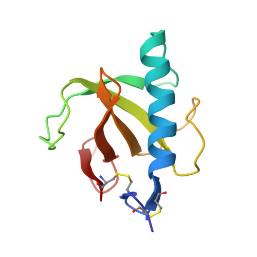
Ribonuclease T1 was purified from an Escherichia coli overproducing strain and co-crystallized with adenosine 2'-monophosphate (2'-AMP) by microdialysis against 50% (v/v) 2-methyl-2,4-pentanediol in 20 mM sodium acetate, 2 mM calcium acetate, pH 4.2. The crystals have orthorhombic space group P2(1)2(1)2(1), with cell dimensions a = 48.93(1), b = 46.57(4), c = 41.04(2) A; Z = 4 and V = 93520 A3. The crystal structure was determined on the basis of the isomorphous structure of uncomplexed RNase T1 (Martinez-Oyanedel et al. (1991) submitted for publication) and refined by least squares methods using stereochemical restraints. The refinement was based on Fhkl of 7,445 reflections with Fo greater than or equal to 1 sigma (Fo) in the resolution range of 10-1.8 A, and converged at a crystallographic R factor of 0.149. The phosphate group of 2'-AMP is tightly hydrogen-bonded to the side chains of the active site residues Tyr38, His40, Glu58, Arg77, and His92, comparable with vanadate binding in the respective complex (Kostrewa, D., Choe, H.-W., Heinemann, U., and Saenger, W. (1989) Biochemistry 28, 7592-7600) and different from the complex with guanosine 2'-monophosphate (Arni, R., Heinemann, U., Tokuoka, R., and Saenger, W. (1988) J. Biol. Chem. 263, 15358-15368) where the phosphate does not interact with Arg77 and His92. The adenosine moiety is not located in the guanosine recognition site but stacked on Gly74 carbonyl and His92 imidazole, which serve as a subsite, as shown previously (Lenz, A., Cordes, F., Heinemann, U., and Saenger, W. (1991) J. Biol. Chem. 266, 7661-7667); in addition, there are hydrogen bonds adenine N6H . . . O Gly74 (minor component of three-center hydrogen bond) and adenosine O5' . . . O delta Asn36. These binding interactions readily explain why RNase T1 has some affinity for 2'-AMP. The molecular structure of RNase T1 is only marginally affected by 2'-AMP binding. Its "empty" guanosine-binding site features a flipped Asn43-Asn44 peptide bond and the side chains of Tyr45, Glu46 adopt conformations typical for RNase T1 not involved in guanosine binding. The side chains of amino acids Leu26, Ser35, Asp49, Val78 are disordered. The disorder of Val78 is of interest since this amino acid is located in a hydrophobic cavity, and the disorder appears to be correlated with an "empty" guanosine-binding site. The two Asp15 carboxylate oxygens and six water molecules coordinate a Ca2+ ion 8-fold in the form of a square antiprism.
Organizational Affiliation:
Institut für Kristallographie, Freie Universität, Berlin, Federal Republic of Germany.