6QSB

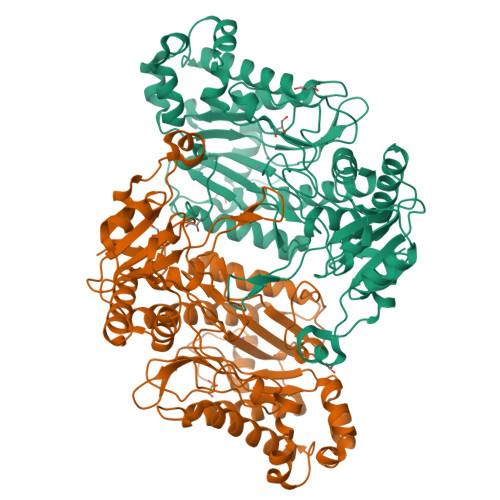
Crystal Structure of Arg470His mutant of Human Prolidase with Mn ions


Experimental Data Snapshot

  • Method: X-RAY DIFFRACTION
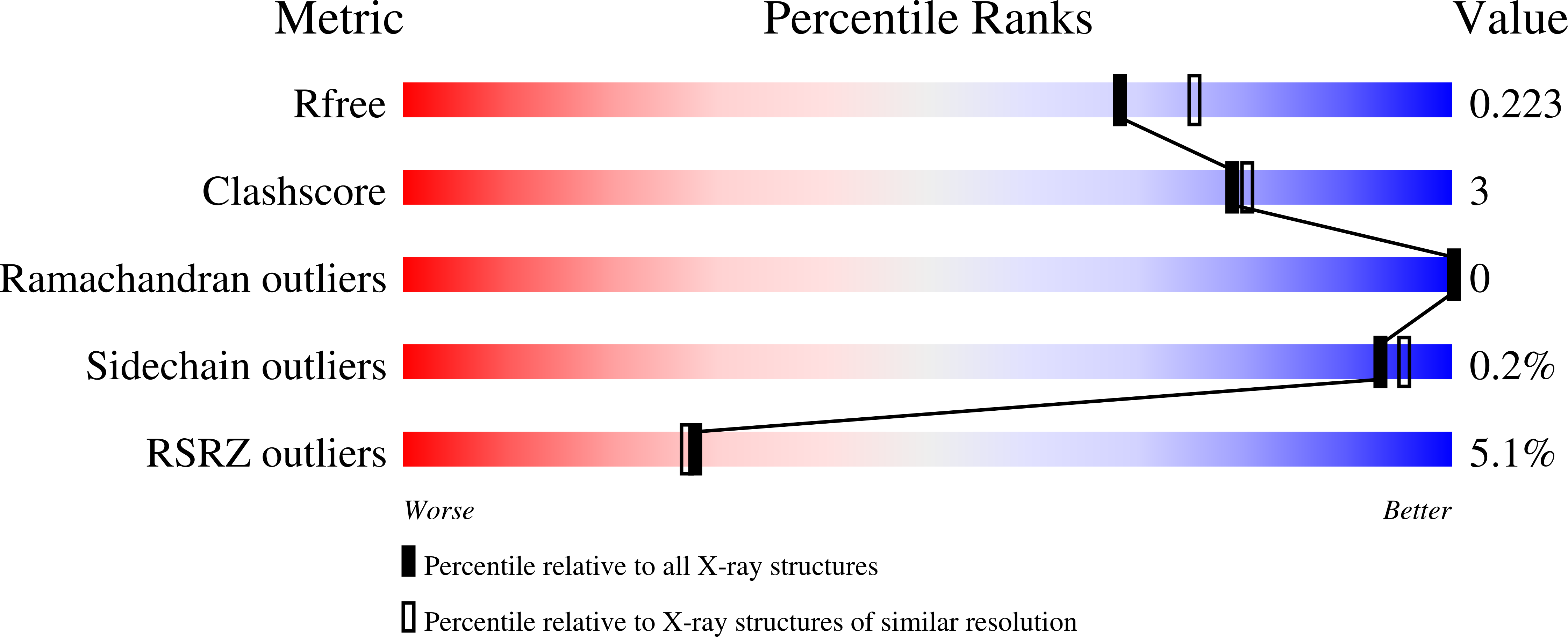
  • Resolution: 1.99 Å
  • R-Value Free: 0.223 
  • R-Value Work: 0.188 
  • R-Value Observed: 0.189 

Starting Model: experimental
View more details

wwPDB Validation   3D Report Full Report


This is version 1.2 of the entry. See complete history


Literature

Structural analysis of new compound heterozygous variants in PEPD gene identified in a patient with Prolidase Deficiency diagnosed by exome sequencing.

Linhares, N.D.Wilk, P.Wator, E.Tostes, M.A.Weiss, M.S.Pena, S.D.J.

(2021) Genet Mol Biol 44: e20200393-e20200393

  • DOI: https://doi.org/10.1590/1678-4685-GMB-2020-0393
  • Primary Citation of Related Structures:  
    6QSB, 6QSC

  • PubMed Abstract: 

    Prolidase Deficiency (PD) is an autosomal recessive rare disorder caused by loss or reduction of prolidase enzymatic activity due to variants in the PEPD gene. PD clinical features vary among affected individuals: skin ulcerations, recurrent infections, and developmental delay are common. In this study, we describe a 16-year-old boy with a mild PD phenotype comprising chronic eczema, recurrent infections and elevated IgE. Whole exome sequencing analysis revealed three PEPD variants: c.575T>C p.(Leu192Pro) inherited from the mother, and c.692_694del p.(Tyr231del) and c.1409G>A p.(Arg470His), both inherited from the father. The variant p.(Tyr231del) has been previously characterized by high-resolution X-ray structure analysis as altering protein dynamics/flexibility. In order to study the effects of the other two prolidase variants, we performed site directed mutagenesis purification and crystallization studies. A high-resolution X-ray structure could only be obtained for the p.(Arg470His) variant, which showed no significant structural differences in comparison to WT prolidase. On the other hand, the p.(Leu192Pro) variant led to significant protein destabilization. Hence, we conclude that the maternal p.(Leu192Pro) variant was likely causally associated with the proband´s disease, together with the known pathogenic paternal variant p.(Tyr231del). Our results demonstrated the utility of exome sequencing to perform diagnosis in PD cases with mild phenotype.


  • Organizational Affiliation

    Universidade Federal de Minas Gerais, Faculdade de Medicina, Laboratório de Genômica Clínica, Belo Horizonte, MG, Brazil.


Macromolecules
Find similar proteins by:  (by identity cutoff)  |  3D Structure
Entity ID: 1
MoleculeChains Sequence LengthOrganismDetailsImage
Xaa-Pro dipeptidase
A, B
493Homo sapiensMutation(s): 1 
Gene Names: PEPDPRD
EC: 3.4.13.9
UniProt & NIH Common Fund Data Resources
Find proteins for P12955 (Homo sapiens)
Explore P12955 
Go to UniProtKB:  P12955
PHAROS:  P12955
GTEx:  ENSG00000124299 
Entity Groups  
Sequence Clusters30% Identity50% Identity70% Identity90% Identity95% Identity100% Identity
UniProt GroupP12955
Sequence Annotations
Expand
  • Reference Sequence
Small Molecules
Ligands 2 Unique
IDChains Name / Formula / InChI Key2D Diagram3D Interactions
GOL
Query on GOL

Download Ideal Coordinates CCD File 
E [auth A]
F [auth A]
G [auth A]
J [auth B]
K [auth B]
E [auth A],
F [auth A],
G [auth A],
J [auth B],
K [auth B],
L [auth B],
M [auth B]
GLYCEROL
C3 H8 O3
PEDCQBHIVMGVHV-UHFFFAOYSA-N
MN
Query on MN

Download Ideal Coordinates CCD File 
C [auth A],
D [auth A],
H [auth B],
I [auth B]
MANGANESE (II) ION
Mn
WAEMQWOKJMHJLA-UHFFFAOYSA-N
Experimental Data & Validation

Experimental Data

Unit Cell:
Length ( Å )Angle ( ˚ )
a = 103.555α = 90
b = 108.533β = 90
c = 211.804γ = 90
Software Package:
Software NamePurpose
XDSdata reduction
PHASERphasing
PHENIXrefinement
PDB_EXTRACTdata extraction
XDSdata scaling

Structure Validation

View Full Validation Report


Entry History 

Deposition Data

Revision History  (Full details and data files)

  • Version 1.0: 2020-03-18
    Type: Initial release
  • Version 1.1: 2024-01-24
    Changes: Data collection, Database references, Derived calculations, Refinement description
  • Version 1.2: 2024-03-06
    Changes: Database references