6CE8

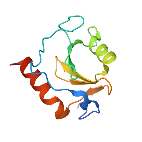
Crystal structure of fragment 2-(Benzo[d]thiazol-2-yl)acetic acid bound in the ubiquitin binding pocket of the HDAC6 zinc-finger domain


Experimental Data Snapshot

  • Method: X-RAY DIFFRACTION
  • Resolution: 1.55 Å
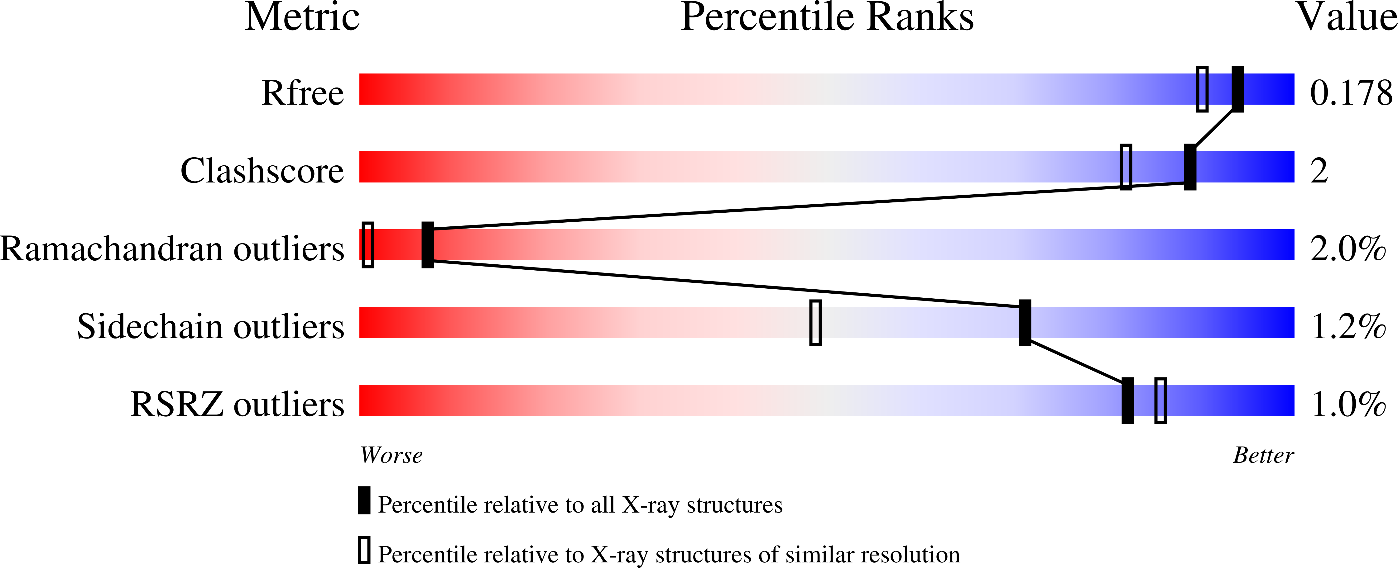
  • R-Value Free: 0.168 
  • R-Value Work: 0.148 
  • R-Value Observed: 0.149 

Starting Model: experimental
View more details

wwPDB Validation   3D Report Full Report


Ligand Structure Quality Assessment 


This is version 1.3 of the entry. See complete history


Literature

Identification and Structure-Activity Relationship of HDAC6 Zinc-Finger Ubiquitin Binding Domain Inhibitors.

Ferreira de Freitas, R.Harding, R.J.Franzoni, I.Ravichandran, M.Mann, M.K.Ouyang, H.Lautens, M.Santhakumar, V.Arrowsmith, C.H.Schapira, M.

(2018) J Med Chem 61: 4517-4527

  • DOI: https://doi.org/10.1021/acs.jmedchem.8b00258
  • Primary Citation of Related Structures:  
    6CE6, 6CE8, 6CEA, 6CEC, 6CED, 6CEE, 6CEF

  • PubMed Abstract: 

    HDAC6 plays a central role in the recruitment of protein aggregates for lysosomal degradation and is a promising target for combination therapy with proteasome inhibitors in multiple myeloma. Pharmacologically displacing ubiquitin from the zinc-finger ubiquitin-binding domain (ZnF-UBD) of HDAC6 is an underexplored alternative to catalytic inhibition. Here, we present the discovery of an HDAC6 ZnF-UBD-focused chemical series and its progression from virtual screening hits to low micromolar inhibitors. A carboxylate mimicking the C-terminal extremity of ubiquitin, and an extended aromatic system stacking with W1182 and R1155, are necessary for activity. One of the compounds induced a conformational remodeling of the binding site where the primary binding pocket opens up onto a ligand-able secondary pocket that may be exploited to increase potency. The preliminary structure-activity relationship accompanied by nine crystal structures should enable further optimization into a chemical probe to investigate the merit of targeting the ZnF-UBD of HDAC6 in multiple myeloma and other diseases.


  • Organizational Affiliation

    Structural Genomics Consortium , University of Toronto , MaRS South Tower, Suite 700, 101 College Street , Toronto , Ontario M5G 1L7 , Canada.


Macromolecules
Find similar proteins by:  (by identity cutoff)  |  3D Structure
Entity ID: 1
MoleculeChains Sequence LengthOrganismDetailsImage
Histone deacetylase 6107Homo sapiensMutation(s): 0 
Gene Names: HDAC6KIAA0901JM21
EC: 3.5.1.98 (PDB Primary Data), 3.5.1 (UniProt)
UniProt & NIH Common Fund Data Resources
Find proteins for Q9UBN7 (Homo sapiens)
Explore Q9UBN7 
Go to UniProtKB:  Q9UBN7
PHAROS:  Q9UBN7
GTEx:  ENSG00000094631 
Entity Groups  
Sequence Clusters30% Identity50% Identity70% Identity90% Identity95% Identity100% Identity
UniProt GroupQ9UBN7
Sequence Annotations
Expand
  • Reference Sequence
Small Molecules
Ligands 3 Unique
IDChains Name / Formula / InChI Key2D Diagram3D Interactions
EYV
Query on EYV

Download Ideal Coordinates CCD File 
I [auth A](1,3-benzothiazol-2-yl)acetic acid
C9 H7 N O2 S
ZOAYQTSFMDZTQA-UHFFFAOYSA-N
ZN
Query on ZN

Download Ideal Coordinates CCD File 
B [auth A],
C [auth A],
D [auth A]
ZINC ION
Zn
PTFCDOFLOPIGGS-UHFFFAOYSA-N
UNX
Query on UNX

Download Ideal Coordinates CCD File 
E [auth A],
F [auth A],
G [auth A],
H [auth A]
UNKNOWN ATOM OR ION
X
Experimental Data & Validation

Experimental Data

Unit Cell:
Length ( Å )Angle ( ˚ )
a = 40.66α = 90
b = 44.41β = 90
c = 55.85γ = 90
Software Package:
Software NamePurpose
REFMACrefinement
Aimlessdata scaling
PDB_EXTRACTdata extraction
xia2data reduction

Structure Validation

View Full Validation Report


Ligand Structure Quality Assessment 


Entry History 

Revision History  (Full details and data files)

  • Version 1.0: 2018-02-28
    Type: Initial release
  • Version 1.1: 2018-05-16
    Changes: Data collection, Database references
  • Version 1.2: 2018-05-30
    Changes: Data collection, Database references
  • Version 1.3: 2023-10-04
    Changes: Data collection, Database references, Refinement description