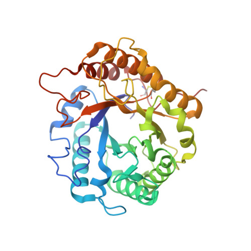
5LJF

Crystal structure of the endo-1,4-glucanase RBcel1 E135A with cellotriose


Experimental Data Snapshot

  • Method: X-RAY DIFFRACTION
  • Resolution: 1.73 Å
  • R-Value Free: 0.212 
  • R-Value Work: 0.176 
  • R-Value Observed: 0.177 

Starting Model: experimental
View more details

wwPDB Validation   3D Report Full Report


This is version 3.2 of the entry. See complete history


Literature

Glycoside hydrolase family 5: structural snapshots highlighting the involvement of two conserved residues in catalysis.

Collet, L.Vander Wauven, C.Oudjama, Y.Galleni, M.Dutoit, R.

(2021) Acta Crystallogr D Struct Biol 77: 205-216

  • DOI: https://doi.org/10.1107/S2059798320015557
  • Primary Citation of Related Structures:  
    5LJF, 6ZZ3

  • PubMed Abstract: 

    The ability of retaining glycoside hydrolases (GHs) to transglycosylate is inherent to the double-displacement mechanism. Studying reaction intermediates, such as the glycosyl-enzyme intermediate (GEI) and the Michaelis complex, could provide valuable information to better understand the molecular factors governing the catalytic mechanism. Here, the GEI structure of RBcel1, an endo-1,4-β-glucanase of the GH5 family endowed with transglycosylase activity, is reported. It is the first structure of a GH5 enzyme covalently bound to a natural oligosaccharide with the two catalytic glutamate residues present. The structure of the variant RBcel1_E135A in complex with cellotriose is also reported, allowing a description of the entire binding cleft of RBcel1. Taken together, the structures deliver different snapshots of the double-displacement mechanism. The structural analysis revealed a significant movement of the nucleophilic glutamate residue during the reaction. Enzymatic assays indicated that, as expected, the acid/base glutamate residue is crucial for the glycosylation step and partly contributes to deglycosylation. Moreover, a conserved tyrosine residue in the -1 subsite, Tyr201, plays a determinant role in both the glycosylation and deglycosylation steps, since the GEI was trapped in the RBcel1_Y201F variant. The approach used to obtain the GEI presented here could easily be transposed to other retaining GHs in clan GH-A.


  • Organizational Affiliation

    LABIRIS, 1 Avenue Emile Gryzon, 1070 Brussels, Belgium.


Macromolecules
Find similar proteins by:  (by identity cutoff)  |  3D Structure
Entity ID: 1
MoleculeChains Sequence LengthOrganismDetailsImage
Endoglucanase
A, B
321uncultured bacteriumMutation(s): 1 
EC: 3.2.1.4
UniProt
Find proteins for C1JI15 (uncultured bacterium)
Explore C1JI15 
Go to UniProtKB:  C1JI15
Entity Groups  
Sequence Clusters30% Identity50% Identity70% Identity90% Identity95% Identity100% Identity
UniProt GroupC1JI15
Sequence Annotations
Expand
  • Reference Sequence
Oligosaccharides

Help

Entity ID: 2
MoleculeChains Length2D Diagram Glycosylation3D Interactions
beta-D-glucopyranose-(1-4)-beta-D-glucopyranose-(1-4)-beta-D-glucopyranose
C, D
3N/A
Glycosylation Resources
GlyTouCan:  G14338VK
GlyCosmos:  G14338VK
GlyGen:  G14338VK
Biologically Interesting Molecules (External Reference) 1 Unique
Experimental Data & Validation

Experimental Data

  • Method: X-RAY DIFFRACTION
  • Resolution: 1.73 Å
  • R-Value Free: 0.212 
  • R-Value Work: 0.176 
  • R-Value Observed: 0.177 
  • Space Group: P 21 21 21
Unit Cell:
Length ( Å )Angle ( ˚ )
a = 45.69α = 90
b = 99.31β = 90
c = 148.58γ = 90
Software Package:
Software NamePurpose
XDSdata reduction
XSCALEdata scaling
PHASERphasing
PHENIXmodel building
Cootmodel building
PHENIXrefinement

Structure Validation

View Full Validation Report


Entry History 

Deposition Data

Revision History  (Full details and data files)

  • Version 1.0: 2017-08-02
    Type: Initial release
  • Version 2.0: 2020-07-29
    Type: Remediation
    Reason: Carbohydrate remediation
    Changes: Advisory, Atomic model, Data collection, Derived calculations, Non-polymer description, Structure summary
  • Version 3.0: 2021-07-21
    Changes: Advisory, Atomic model, Data collection, Database references, Derived calculations, Structure summary
  • Version 3.1: 2024-01-10
    Changes: Data collection, Database references, Refinement description
  • Version 3.2: 2024-10-16
    Changes: Structure summary