5EB3

VB6-bound protein


Experimental Data Snapshot

  • Method: X-RAY DIFFRACTION
  • Resolution: 2.40 Å
  • R-Value Free: 0.198 
  • R-Value Work: 0.178 
  • R-Value Observed: 0.179 

Starting Model: experimental
View more details

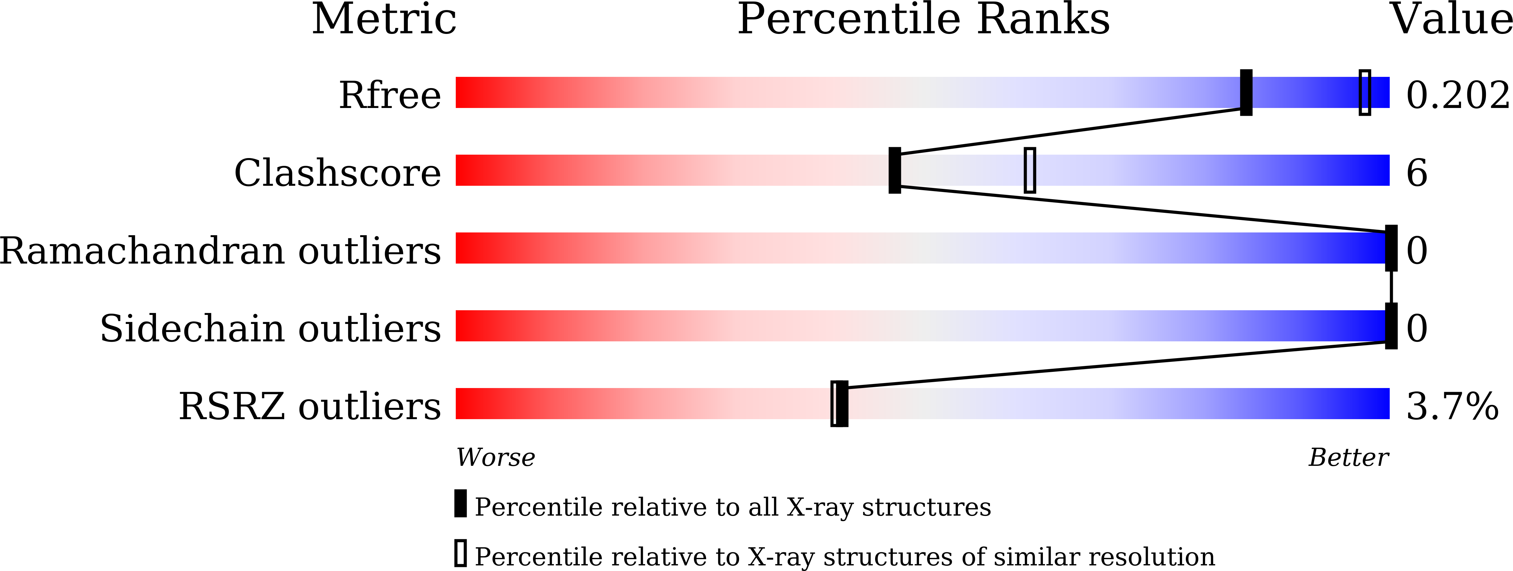
wwPDB Validation   3D Report Full Report


Ligand Structure Quality Assessment 


This is version 1.3 of the entry. See complete history


Literature

Structural insights into the regulatory mechanism of the Pseudomonas aeruginosa YfiBNR system

Xu, M.Yang, X.Yang, X.-A.Zhou, L.Liu, T.-Z.Fan, Z.Jiang, T.

(2016) Protein Cell 7: 403-416

  • DOI: https://doi.org/10.1007/s13238-016-0264-7
  • Primary Citation of Related Structures:  
    5EAZ, 5EB0, 5EB1, 5EB2, 5EB3

  • PubMed Abstract: 

    YfiBNR is a recently identified bis-(3'-5')-cyclic dimeric GMP (c-di-GMP) signaling system in opportunistic pathogens. It is a key regulator of biofilm formation, which is correlated with prolonged persistence of infection and antibiotic drug resistance. In response to cell stress, YfiB in the outer membrane can sequester the periplasmic protein YfiR, releasing its inhibition of YfiN on the inner membrane and thus provoking the diguanylate cyclase activity of YfiN to induce c-di-GMP production. However, the detailed regulatory mechanism remains elusive. Here, we report the crystal structures of YfiB alone and of an active mutant YfiB(L43P) complexed with YfiR with 2:2 stoichiometry. Structural analyses revealed that in contrast to the compact conformation of the dimeric YfiB alone, YfiB(L43P) adopts a stretched conformation allowing activated YfiB to penetrate the peptidoglycan (PG) layer and access YfiR. YfiB(L43P) shows a more compact PG-binding pocket and much higher PG binding affinity than wild-type YfiB, suggesting a tight correlation between PG binding and YfiB activation. In addition, our crystallographic analyses revealed that YfiR binds Vitamin B6 (VB6) or L-Trp at a YfiB-binding site and that both VB6 and L-Trp are able to reduce YfiB(L43P)-induced biofilm formation. Based on the structural and biochemical data, we propose an updated regulatory model of the YfiBNR system.


  • Organizational Affiliation

    National Laboratory of Biomacromolecules, Institute of Biophysics, Chinese Academy of Sciences, Beijing, 100101, China.


Macromolecules
Find similar proteins by:  (by identity cutoff)  |  3D Structure
Entity ID: 1
MoleculeChains Sequence LengthOrganismDetailsImage
YfiR
A, B
159Pseudomonas aeruginosa PAO1Mutation(s): 0 
Gene Names: yfiRPA1121
UniProt
Find proteins for Q9I4L4 (Pseudomonas aeruginosa (strain ATCC 15692 / DSM 22644 / CIP 104116 / JCM 14847 / LMG 12228 / 1C / PRS 101 / PAO1))
Explore Q9I4L4 
Go to UniProtKB:  Q9I4L4
Entity Groups  
Sequence Clusters30% Identity50% Identity70% Identity90% Identity95% Identity100% Identity
UniProt GroupQ9I4L4
Sequence Annotations
Expand
  • Reference Sequence
Experimental Data & Validation

Experimental Data

  • Method: X-RAY DIFFRACTION
  • Resolution: 2.40 Å
  • R-Value Free: 0.198 
  • R-Value Work: 0.178 
  • R-Value Observed: 0.179 
  • Space Group: P 43 21 2
Unit Cell:
Length ( Å )Angle ( ˚ )
a = 120.236α = 90
b = 120.236β = 90
c = 84.993γ = 90
Software Package:
Software NamePurpose
PHENIXrefinement
HKL-2000data scaling
PDB_EXTRACTdata extraction
d*TREKdata reduction
PHASERphasing

Structure Validation

View Full Validation Report


Ligand Structure Quality Assessment 


Entry History & Funding Information

Deposition Data


Funding OrganizationLocationGrant Number
National Natural Science FoundationChina31570768
National Natural Science FoundationChina91419308

Revision History  (Full details and data files)

  • Version 1.0: 2016-05-18
    Type: Initial release
  • Version 1.1: 2016-06-15
    Changes: Database references
  • Version 1.2: 2023-11-08
    Changes: Data collection, Database references, Derived calculations, Refinement description
  • Version 1.3: 2024-11-13
    Changes: Structure summary