


Crystal structure of SmyD3 in complex with covalent inhibitor 1
Baburajendran, N., Anna E, J.To be published.
Experimental Data Snapshot
Starting Model: experimental
View more details
wwPDB Validation 3D Report Full Report

Entity ID: 1 | |||||
|---|---|---|---|---|---|
| Molecule | Chains | Sequence Length | Organism | Details | Image |
| Smyd3 methyltransferase | 423 | Homo sapiens | Mutation(s): 0 | 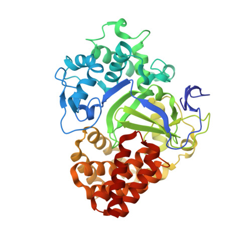 | |
UniProt & NIH Common Fund Data Resources | |||||
Find proteins for Q9H7B4 (Homo sapiens) Explore Q9H7B4 Go to UniProtKB: Q9H7B4 | |||||
PHAROS: Q9H7B4 GTEx: ENSG00000185420 | |||||
Entity Groups | |||||
| Sequence Clusters | 30% Identity50% Identity70% Identity90% Identity95% Identity100% Identity | ||||
| UniProt Group | Q9H7B4 | ||||
Sequence AnnotationsExpand | |||||
| |||||
| Ligands 3 Unique | |||||
|---|---|---|---|---|---|
| ID | Chains | Name / Formula / InChI Key | 2D Diagram | 3D Interactions | |
| SAM Query on SAM | B [auth A] | S-ADENOSYLMETHIONINE C15 H22 N6 O5 S MEFKEPWMEQBLKI-FCKMPRQPSA-N |  | ||
| 8NR Query on 8NR | F [auth A] | ethyl 4-(5,6,7,8-tetrahydroacridin-3-ylcarbonyl)piperazine-1-carboxylate C21 H25 N3 O3 YMYFCDFYTNCCDJ-UHFFFAOYSA-N |  | ||
| ZN Query on ZN | C [auth A], D [auth A], E [auth A] | ZINC ION Zn PTFCDOFLOPIGGS-UHFFFAOYSA-N |  | ||
| Length ( Å ) | Angle ( ˚ ) |
|---|---|
| a = 61.458 | α = 90 |
| b = 66.472 | β = 90 |
| c = 107.062 | γ = 90 |
| Software Name | Purpose |
|---|---|
| PHENIX | refinement |
| SCALA | data scaling |
| PDB_EXTRACT | data extraction |
RCSB PDB (citation) is hosted by
RCSB PDB is a member of the

