5RNT

X-RAY ANALYSIS OF CUBIC CRYSTALS OF THE COMPLEX FORMED BETWEEN RIBONUCLEASE T1 AND GUANOSINE-3',5'-BISPHOSPHATE


Experimental Data Snapshot

  • Method: X-RAY DIFFRACTION
  • Resolution: 3.20 Å
  • R-Value Observed: 0.166 

wwPDB Validation   3D Report Full Report


Ligand Structure Quality Assessment 


This is version 1.3 of the entry. See complete history

Re-refinement Note

A newer entry is available that reflects an alternative modeling of the original data: 3URP


Literature

X-ray analysis of cubic crystals of the complex formed between ribonuclease T1 and guanosine-3',5'-bisphosphate.

Lenz, A.Heinemann, U.Maslowska, M.Saenger, W.

(1991) Acta Crystallogr B 47: 521-527

  • DOI: https://doi.org/10.1107/s0108768191001684
  • Primary Citation of Related Structures:  
    5RNT

  • PubMed Abstract: 

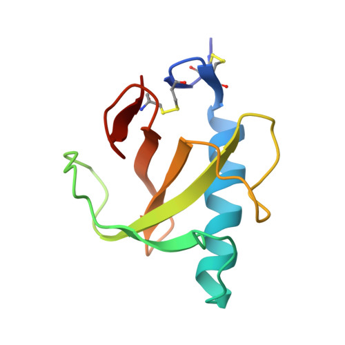
    The complex formed between ribonuclease T1 (RNase T1) and guanosine-3',5'-bisphosphate (3',5'-pGp) crystallizes in the cubic space group I23 with alpha = 86.47 (4) A. X-ray data were collected on a four-circle diffractometer to 3.2 A resolution and the structure was determined by molecular-replacement methods [ULTIMA; Rabinovich & Shakked (1984). Acta Cryst. A40, 195-200] based on the RNase T1 coordinates taken from the complex with guanosine-2'-phosphate. Refinement converged at 16.6% for 1540 data with Fo greater than 1 sigma (Fo) with acceptable stereochemistry. The RNase T1 conformation is comparable to that in other complexes which crystallize preferentially in space group P2(1)2(1)2(1) except for side chains that interact intermolecularly. The guanine of 3',5'-pGp is bound to the recognition site in the same way as in other guanine-containing complexes except for its interaction with Glu46. The side-chain carboxylate of this amino acid does not form hydrogen bonds to N1H and N2H of guanine but is rotated so as to permit insertion of two water molecules which replace its acceptor functions. In contrast to other guanosine derivatives which are bound to RNase T1 in the syn form, 3',5'-pGp is anti. This conformation positions the two phosphate groups 'outside' the protein, with hydrogen-bonding contacts only to water molecules; the active site is filled by water. The RNase T1-3',5'-pGp complex probably has biological significance as it may represent the enzyme-product complex before dissociation.


  • Organizational Affiliation

    Institut für Kristallographie, Freie Universität Berlin, Germany.


Macromolecules
Find similar proteins by:  (by identity cutoff)  |  3D Structure
Entity ID: 1
MoleculeChains Sequence LengthOrganismDetailsImage
RIBONUCLEASE T1104Aspergillus oryzaeMutation(s): 0 
EC: 3.1.27.3
UniProt
Find proteins for P00651 (Aspergillus oryzae (strain ATCC 42149 / RIB 40))
Explore P00651 
Go to UniProtKB:  P00651
Entity Groups  
Sequence Clusters30% Identity50% Identity70% Identity90% Identity95% Identity100% Identity
UniProt GroupP00651
Sequence Annotations
Expand
  • Reference Sequence
Small Molecules
Ligands 1 Unique
IDChains Name / Formula / InChI Key2D Diagram3D Interactions
PGP
Query on PGP

Download Ideal Coordinates CCD File 
B [auth A]GUANOSINE-3',5'-DIPHOSPHATE
C10 H15 N5 O11 P2
XYVNHPYNSPGYLI-UUOKFMHZSA-N
Experimental Data & Validation

Experimental Data

  • Method: X-RAY DIFFRACTION
  • Resolution: 3.20 Å
  • R-Value Observed: 0.166 
  • Space Group: I 2 3
Unit Cell:
Length ( Å )Angle ( ˚ )
a = 86.47α = 90
b = 86.47β = 90
c = 86.47γ = 90
Software Package:
Software NamePurpose
PROFFTrefinement

Structure Validation

View Full Validation Report


Ligand Structure Quality Assessment 


Entry History 

Deposition Data

Revision History  (Full details and data files)

  • Version 1.0: 1993-01-15
    Type: Initial release
  • Version 1.1: 2008-03-25
    Changes: Version format compliance
  • Version 1.2: 2011-07-13
    Changes: Version format compliance
  • Version 1.3: 2017-11-29
    Changes: Derived calculations, Other