5HXB

Cereblon in complex with DDB1, CC-885, and GSPT1


Experimental Data Snapshot

  • Method: X-RAY DIFFRACTION
  • Resolution: 3.60 Å
  • R-Value Free: 0.273 
  • R-Value Work: 0.224 
  • R-Value Observed: 0.226 

Starting Models: experimental
View more details

wwPDB Validation   3D Report Full Report


Ligand Structure Quality Assessment 


This is version 1.5 of the entry. See complete history


Literature

A novel cereblon modulator recruits GSPT1 to the CRL4(CRBN) ubiquitin ligase.

Matyskiela, M.E.Lu, G.Ito, T.Pagarigan, B.Lu, C.C.Miller, K.Fang, W.Wang, N.Y.Nguyen, D.Houston, J.Carmel, G.Tran, T.Riley, M.Nosaka, L.Lander, G.C.Gaidarova, S.Xu, S.Ruchelman, A.L.Handa, H.Carmichael, J.Daniel, T.O.Cathers, B.E.Lopez-Girona, A.Chamberlain, P.P.

(2016) Nature 535: 252-257

  • DOI: https://doi.org/10.1038/nature18611
  • Primary Citation of Related Structures:  
    5HXB

  • PubMed Abstract: 

    Immunomodulatory drugs bind to cereblon (CRBN) to confer differentiated substrate specificity on the CRL4(CRBN) E3 ubiquitin ligase. Here we report the identification of a new cereblon modulator, CC-885, with potent anti-tumour activity. The anti-tumour activity of CC-885 is mediated through the cereblon-dependent ubiquitination and degradation of the translation termination factor GSPT1. Patient-derived acute myeloid leukaemia tumour cells exhibit high sensitivity to CC-885, indicating the clinical potential of this mechanism. Crystallographic studies of the CRBN-DDB1-CC-885-GSPT1 complex reveal that GSPT1 binds to cereblon through a surface turn containing a glycine residue at a key position, interacting with both CC-885 and a 'hotspot' on the cereblon surface. Although GSPT1 possesses no obvious structural, sequence or functional homology to previously known cereblon substrates, mutational analysis and modelling indicate that the cereblon substrate Ikaros uses a similar structural feature to bind cereblon, suggesting a common motif for substrate recruitment. These findings define a structural degron underlying cereblon 'neosubstrate' selectivity, and identify an anti-tumour target rendered druggable by cereblon modulation.


  • Organizational Affiliation

    Celgene Corporation, 10300 Campus Point Drive, Suite 100, San Diego, California 92121, USA.


Macromolecules
Find similar proteins by:  (by identity cutoff)  |  3D Structure
Entity ID: 1
MoleculeChains Sequence LengthOrganismDetailsImage
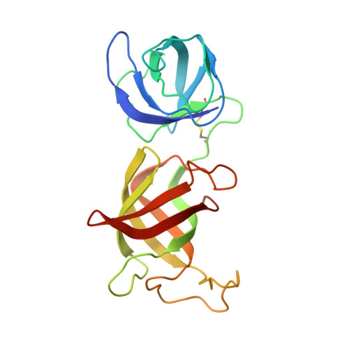
Eukaryotic peptide chain release factor GTP-binding subunit ERF3AA [auth X],
F [auth A]
199Homo sapiensMutation(s): 0 
Gene Names: GSPT1ERF3A
EC: 3.6.5
UniProt & NIH Common Fund Data Resources
Find proteins for P15170 (Homo sapiens)
Explore P15170 
Go to UniProtKB:  P15170
PHAROS:  P15170
GTEx:  ENSG00000103342 
Entity Groups  
Sequence Clusters30% Identity50% Identity70% Identity90% Identity95% Identity100% Identity
UniProt GroupP15170
Sequence Annotations
Expand
  • Reference Sequence
Find similar proteins by:  (by identity cutoff)  |  3D Structure
Entity ID: 2
MoleculeChains Sequence LengthOrganismDetailsImage
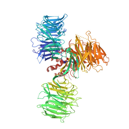
DNA damage-binding protein 1B [auth Y],
D [auth B]
1,140Homo sapiensMutation(s): 0 
Gene Names: DDB1XAP1
UniProt & NIH Common Fund Data Resources
Find proteins for Q16531 (Homo sapiens)
Explore Q16531 
Go to UniProtKB:  Q16531
PHAROS:  Q16531
GTEx:  ENSG00000167986 
Entity Groups  
Sequence Clusters30% Identity50% Identity70% Identity90% Identity95% Identity100% Identity
UniProt GroupQ16531
Sequence Annotations
Expand
  • Reference Sequence
Find similar proteins by:  (by identity cutoff)  |  3D Structure
Entity ID: 3
MoleculeChains Sequence LengthOrganismDetailsImage
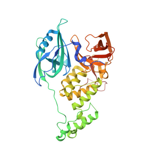
Protein cereblonC [auth Z],
E [auth C]
406Homo sapiensMutation(s): 0 
Gene Names: CRBNAD-006
UniProt & NIH Common Fund Data Resources
Find proteins for Q96SW2 (Homo sapiens)
Explore Q96SW2 
Go to UniProtKB:  Q96SW2
PHAROS:  Q96SW2
GTEx:  ENSG00000113851 
Entity Groups  
Sequence Clusters30% Identity50% Identity70% Identity90% Identity95% Identity100% Identity
UniProt GroupQ96SW2
Sequence Annotations
Expand
  • Reference Sequence
Experimental Data & Validation

Experimental Data

  • Method: X-RAY DIFFRACTION
  • Resolution: 3.60 Å
  • R-Value Free: 0.273 
  • R-Value Work: 0.224 
  • R-Value Observed: 0.226 
  • Space Group: P 1 2 1
Unit Cell:
Length ( Å )Angle ( ˚ )
a = 156.747α = 90
b = 111.522β = 95.81
c = 175.057γ = 90
Software Package:
Software NamePurpose
REFMACrefinement
HKL-2000data reduction
SCALEPACKdata scaling
PHASERphasing

Structure Validation

View Full Validation Report


Ligand Structure Quality Assessment 


Entry History 

Deposition Data

Revision History  (Full details and data files)

  • Version 1.0: 2016-06-29
    Type: Initial release
  • Version 1.1: 2016-07-06
    Changes: Database references
  • Version 1.2: 2016-07-27
    Changes: Database references
  • Version 1.3: 2017-11-01
    Changes: Advisory, Author supporting evidence, Database references, Derived calculations
  • Version 1.4: 2023-09-27
    Changes: Data collection, Database references, Refinement description
  • Version 1.5: 2024-10-23
    Changes: Structure summary