4RJD

TFP bound in alternate orientations to calcium-saturated Calmodulin C-Domains


Experimental Data Snapshot

  • Method: X-RAY DIFFRACTION
  • Resolution: 2.00 Å
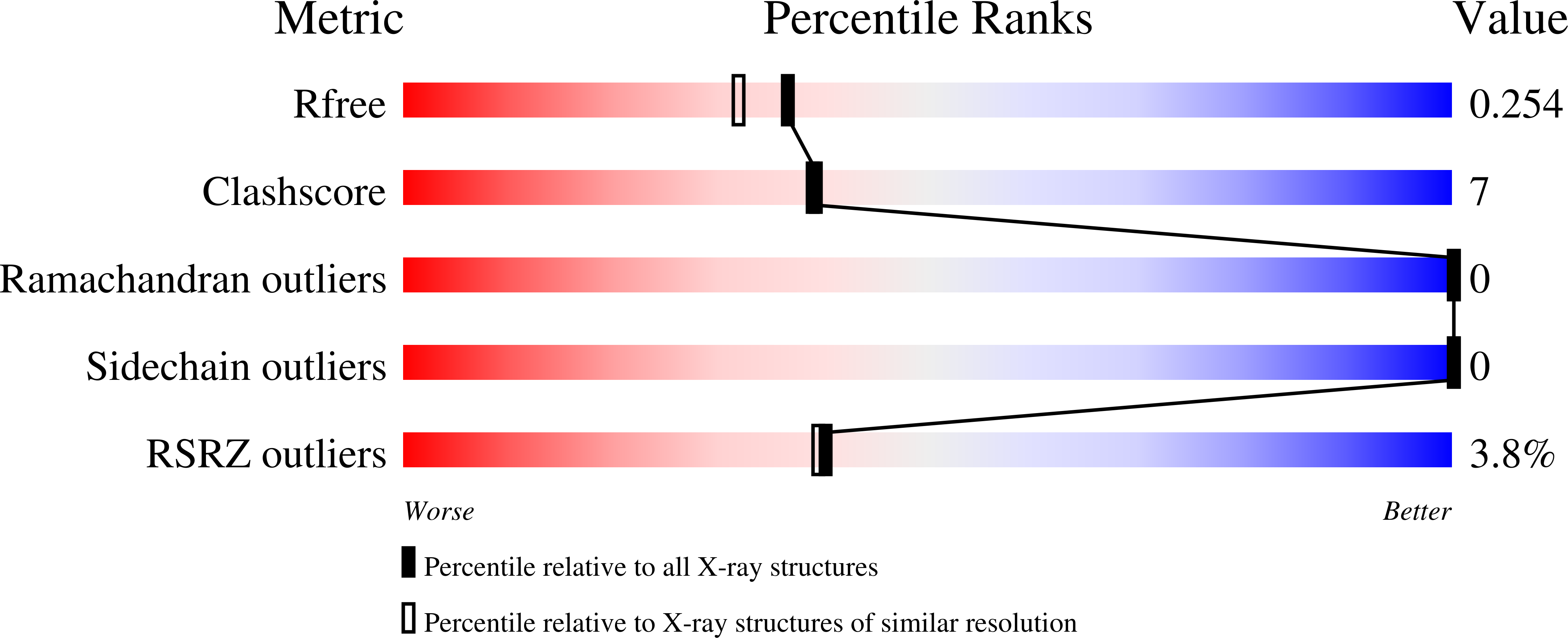
  • R-Value Free: 0.253 
  • R-Value Work: 0.177 
  • R-Value Observed: 0.181 

Starting Model: experimental
View more details

wwPDB Validation   3D Report Full Report


Ligand Structure Quality Assessment 


This is version 1.3 of the entry. See complete history


Literature

Opposing orientations of the anti-psychotic drug trifluoperazine selected by alternate conformations of M144 in calmodulin.

Feldkamp, M.D.Gakhar, L.Pandey, N.Shea, M.A.

(2015) Proteins 83: 989-996

  • DOI: https://doi.org/10.1002/prot.24781
  • Primary Citation of Related Structures:  
    4RJD

  • PubMed Abstract: 

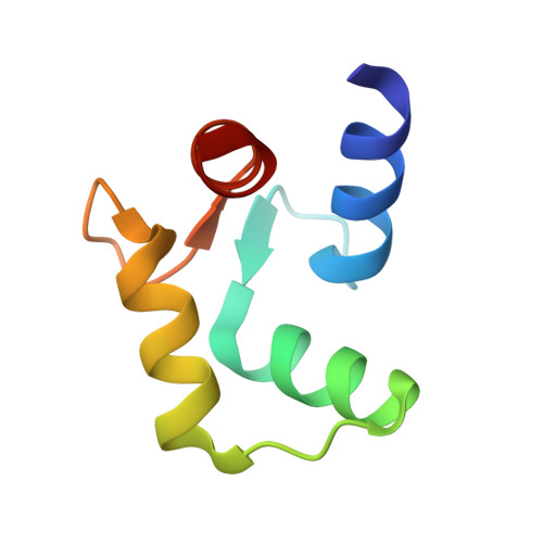
    The anti-psychotic drug trifluoperazine (TFP) is an antagonist observed to bind to calcium-saturated calmodulin ((Ca(2+) )4 -CaM) at ratios of 1:1 (1CTR), 2:1 (1A29), and 4:1 (1LIN). Each structure contains one TFP bound in the hydrophobic cleft of the C-domain of CaM. However, the orientation of the trifluoromethyl (CF3 ) moiety differs among them: it is buried in the C-domain cleft of 1A29 and 1LIN, but protrudes from 1CTR. We report a 2.0 Å resolution crystallographic structure (4RJD) of TFP bound to the (Ca(2+) )-saturated C-domain of CaM (CaMC ). The asymmetric unit contains two molecules of (Ca(2+) )2 -CaMC . Chain backbones were nearly identical, but the orientation of TFP in the cleft of Chain A matched 1A29/1LIN, while TFP bound to Chain B matched 1CTR. This was accommodated by a flip of the M144 sidechain and small changes in sidechains of M109 and M145. Docking simulations suggested that the rotamer conformation of M144 determined the orientation of TFP within the cleft of (Ca(2+) )2 -CaMC . Chains A and B show that the open cleft of (Ca(2+) )2 -CaMC is promiscuous in accepting TFP in reversed directions under the same crystallization conditions. Observing multiple orientations of an antagonist bound to a single protein highlights the challenge of designing highly specific pharmaceuticals, and may have importance for QSAR of other CF3 -containing drugs such as fluoxetine (anti-depressant) or efavirenz (reverse transcriptase inhibitor). This study emphasizes that a single structure of a complex represents an energetically accessible state, but does not necessarily show the full range of energetically equivalent states.


  • Organizational Affiliation

    Department of Biochemistry, Roy J. and Lucille A. Carver College of Medicine, University of Iowa, Iowa City, Iowa, 52242-1109.


Macromolecules
Find similar proteins by:  (by identity cutoff)  |  3D Structure
Entity ID: 1
MoleculeChains Sequence LengthOrganismDetailsImage
Calmodulin
A, B
66Rattus norvegicusMutation(s): 0 
Gene Names: Calm1CalmCamCam1Calm2Cam2CambCalm3Cam3Camc
UniProt
Find proteins for P0DP29 (Rattus norvegicus)
Explore P0DP29 
Go to UniProtKB:  P0DP29
Entity Groups  
Sequence Clusters30% Identity50% Identity70% Identity90% Identity95% Identity100% Identity
UniProt GroupP0DP29
Sequence Annotations
Expand
  • Reference Sequence
Small Molecules
Ligands 6 Unique
IDChains Name / Formula / InChI Key2D Diagram3D Interactions
TFP
Query on TFP

Download Ideal Coordinates CCD File 
E [auth A],
F [auth A],
L [auth B],
M [auth B]
10-[3-(4-METHYL-PIPERAZIN-1-YL)-PROPYL]-2-TRIFLUOROMETHYL-10H-PHENOTHIAZINE
C21 H24 F3 N3 S
ZEWQUBUPAILYHI-UHFFFAOYSA-N
1PE
Query on 1PE

Download Ideal Coordinates CCD File 
G [auth A]PENTAETHYLENE GLYCOL
C10 H22 O6
JLFNLZLINWHATN-UHFFFAOYSA-N
OXL
Query on OXL

Download Ideal Coordinates CCD File 
I [auth A]OXALATE ION
C2 O4
MUBZPKHOEPUJKR-UHFFFAOYSA-L
SCN
Query on SCN

Download Ideal Coordinates CCD File 
N [auth B]THIOCYANATE ION
C N S
ZMZDMBWJUHKJPS-UHFFFAOYSA-M
CA
Query on CA

Download Ideal Coordinates CCD File 
C [auth A],
D [auth A],
J [auth B],
K [auth B]
CALCIUM ION
Ca
BHPQYMZQTOCNFJ-UHFFFAOYSA-N
CL
Query on CL

Download Ideal Coordinates CCD File 
H [auth A],
O [auth B]
CHLORIDE ION
Cl
VEXZGXHMUGYJMC-UHFFFAOYSA-M
Experimental Data & Validation

Experimental Data

  • Method: X-RAY DIFFRACTION
  • Resolution: 2.00 Å
  • R-Value Free: 0.253 
  • R-Value Work: 0.177 
  • R-Value Observed: 0.181 
  • Space Group: P 1 21 1
Unit Cell:
Length ( Å )Angle ( ˚ )
a = 24.606α = 90
b = 85.637β = 93
c = 35.341γ = 90
Software Package:
Software NamePurpose
Blu-Icedata collection
PHASERphasing
PHENIXrefinement
d*TREKdata reduction
d*TREKdata scaling

Structure Validation

View Full Validation Report


Ligand Structure Quality Assessment 


Entry History 

Deposition Data

Revision History  (Full details and data files)

  • Version 1.0: 2015-08-26
    Type: Initial release
  • Version 1.1: 2017-11-22
    Changes: Refinement description
  • Version 1.2: 2020-01-29
    Changes: Derived calculations
  • Version 1.3: 2023-09-20
    Changes: Data collection, Database references, Derived calculations, Refinement description